3221
Automatic brain segmentation framework for bias field rich cranial MRI scans of rats and mice via similarity invariant shape priors1Department of Computer Science, University of Copenhagen, Copenhagen, Denmark, 2Champalimaud Centre for the Unknown, Champalimaud Research, Lisbon, Portugal, 3Center for Translational Neuromedicine, University of Copenhagen, Copenhagen, Denmark, 4Department of Anesthesiology, Yale School of Medicine, Yale University, New Haven, CT, United States, 5Center for Translational Neuromedicine, University of Rochester, Rochester, NY, United States
Synopsis
This abstract presents an extension to our previous work for the extraction of rat brain tissue and internal cerebrospinal fluid networks in MR imaging of rat crania that display severe bias fields. This work contributes automation and robustness in the skull stripping module by introducing an automatic similarity invariant shape prior segmentation method. We demonstrate the capabilities of our framework on both rat brain as well as mouse brain data, using the same minimal number of rat brain priors.
Introduction
MR imaging using surface receiver coils are in general affected by strong image bias fields, for which only partial correction is available. This makes subsequent feature extraction and analysis a difficult task. In a previous work1 we presented a semi-automatic framework that was able to segment and extract rat brain tissue and the cerebrospinal fluid (CSF) networks. In this work we improve on the robustness of the method and make it fully automatic by introducing segmentation shape priors which, by construction, are invariant to rotation, scale, and translation. We show that our improved method obtains very good segmentation results, while automating the process and limiting the number of free parameters.
Animals
Source 1 (identical to our previous work1): Unaffected male rats of the Wistar Kyoto stain (WKY) and spontaneously hypertensive rats (SHR) were obtained from Charles River, Germany. Separate groups of rats were scanned at two age-ranges: young (7-9 weeks old; WKY7, n = 11; and SHR7, n = 8) and young adults (19-21 weeks WKY19, n = 8; and SHR19, n= 9). All treatments and imaging were performed according to protocols approved by the IACUC, and according to a protocol approved by the University of Copenhagen animal experimentation committee.
Source 2: Male C57Bl6 mice (7-8 weeks old) were induced into deep anesthesia with 5% isoflurane and maintained under 2% isoflurane during the MRI experiments. All animal experiments were preapproved by the institutional and national authorities and were carried out according to European Directive 2010/63.
MRI
Source 1: All MRI investigations were conducted on a 9.4 T magnet (Bruker Biospec 9.4/30 USR) interfaced to a Bruker Advance III console and controlled by Paravision 5.1 software (Bruker BioSpin) at the Panum Institute, University of Copenhagen. Imaging was performed with an 86 mm3 volume resonator and a surface quadrature array receiver coil. Images were acquired with the spoiled gradient FLASH3D sequence (TE: 4 ms, TR: 15 ms, NA: 3; matrix: 128 x 128 x 128, voxel size: 0.24 x 0.24 x 0.26 mm, FA: 15°, scan time: 4:05 min).
Source 2: MRI was performed on a 9.4T Bruker BioSpec scanner controlled by Paravision 6.0.1 software, using a 86 mm3 quadrature resonator for transmittance and a 4-element cryoprobe for reception. Images were acquired with a 3D multi-gradient echo sequence (16 echos, TE1: 2.2 ms, 𝚫TE: 2.2 ms , TR: 100 ms , matrix: 180 x 90 x 115, voxel size: 0.11 x 0.1x 0.1 mm, FA: 60°, scan time: 12:16 min). Processing was performed on the mean across all 16 echo times.
Methods
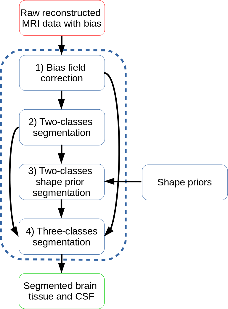
Our pipeline is modified from Hansen et al1. It consists of 1) bias correction, 2) a preliminary 2-classes segmentation, robust to remaining bias, 3) a segmentation with a proposed kernel density shape prior to automatically detect and segment the brain/CSF segments, and 4) a 3-classes (background, brain tissue and CSF) segmentation. Step 1, 2, and 4 are identical to Hansen et al.1.
Brain shape prior segmentation. In this step, we combine a Chan-Vese data term and a similarity invariant shape prior, built from $$$n$$$ training / template shapes. We have only used $$$n=4$$$ training shapes here. We use the following shape prior based segmentation energy
$$\mathcal{E}(v) = \frac12\int_\Omega\left((u-c_1)^2v + (u-c_2)^2(1-v)\right)dx + \mathcal{S}(v)$$
where
$$\mathcal{S}(v) = -\log\sum_{i=1}^{n}e^{-\frac{d^2(v,v_i)}{2\rho^2}}$$
is the contribution from a kernel density estimated collection of training shapes $$$v_i$$$. $$$d^2$$$ is a pseudo squared distance measure.
Results
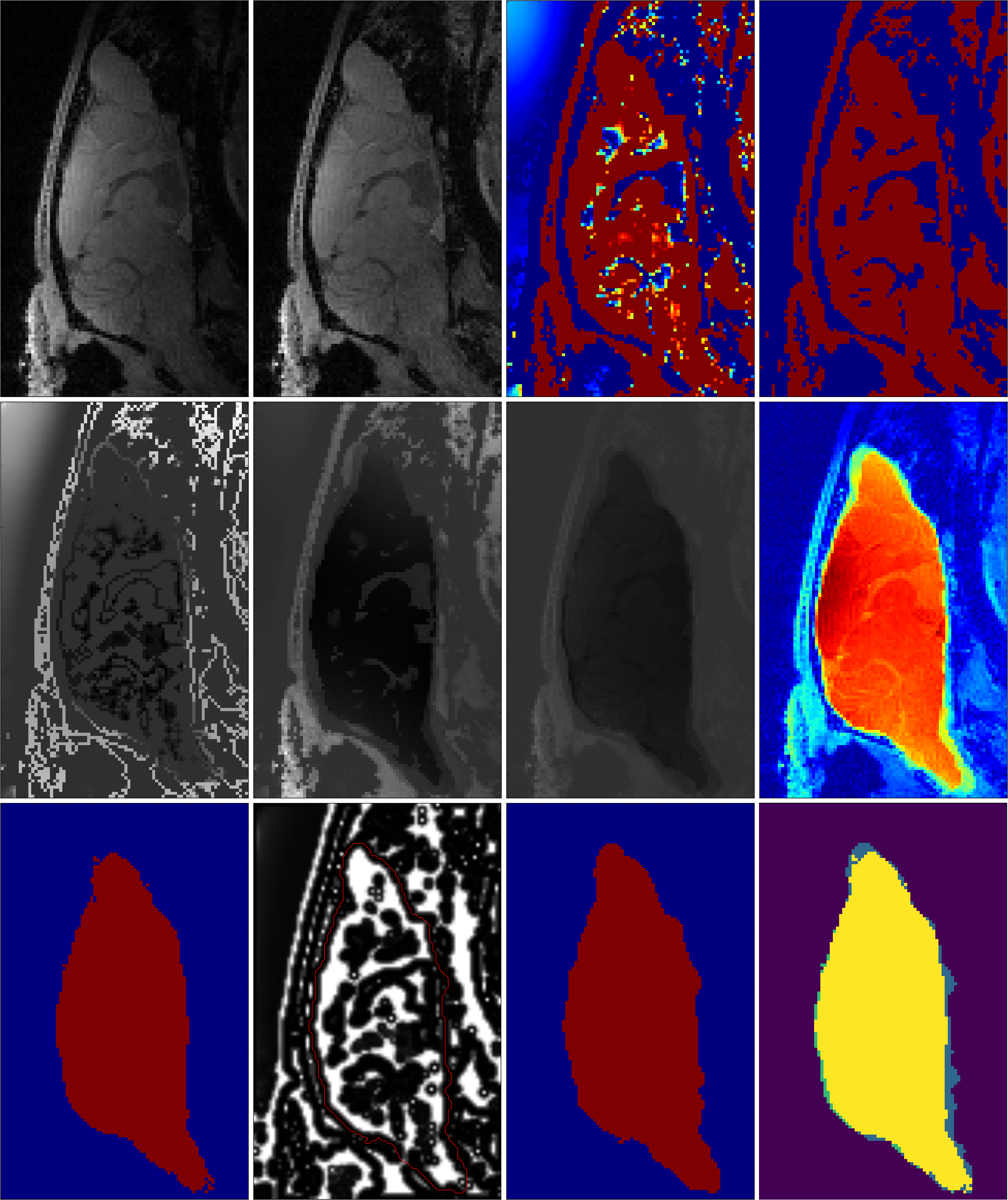
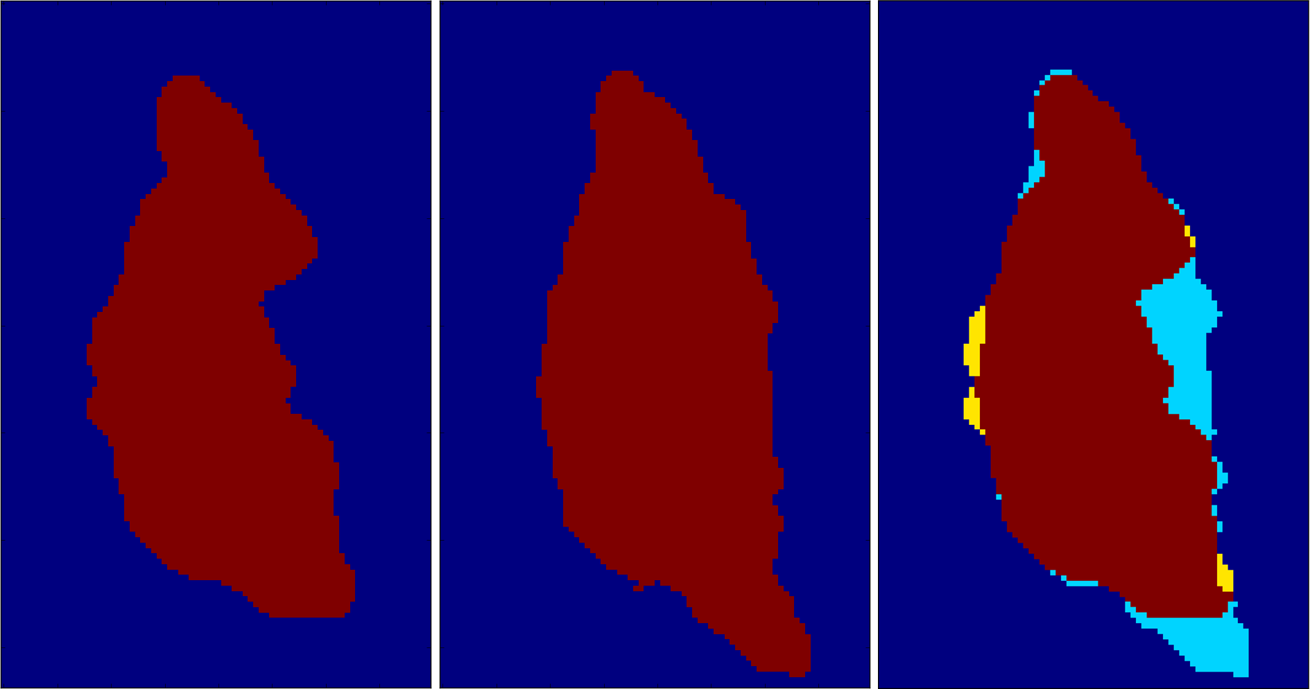
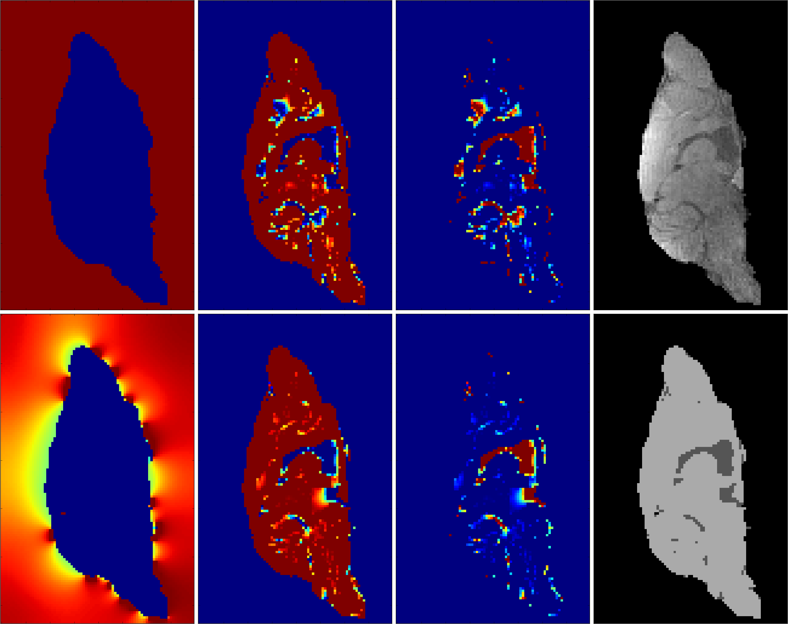
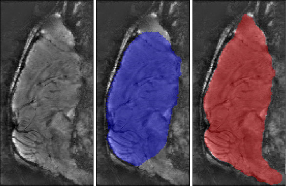
We ran our brain segmentation on rat brain data from source 1 and on mouse data from source 2. The results for step 1-3 are illustrated in Figure 2, while a comparison with our former brain extraction routine can be found in Figure 3, The final segmentation result is shown in Figure 4. Figure 5 highlights our brain extraction method using rat data for training, but running on mouse data and compares it to that of AFNI's 3D skullstrip.Discussion and Conclusion
We have proposed an automatic framework for rat brain and internal CSF network segmentation for images affected by strong bias fields, using a brain shape prior. It is shown to work well for data coming from other sources than the one used for training our shape prior. To improve the results further, extra training shapes representing better shape variability should be incorporated.Acknowledgements
No acknowledgement found.References
1. Hansen et al., Brain extraction and segmentation framework for bias field rich cranial MRI scans of rats, ISMRM 2018, Paris, France.
2. Tustison et al., N4ITK: improved N3 bias correction. IEEE Trans. Med. Im. 2010
3. Hansen, J. D. K., and Lauze, F. Local Mean Multiphase Segmentation with HMMF Models. In Proc. SSVM 2017 (2017), Springer, pp. 396–407.
Figures