3220
Structural changes in a rat model of traumatic brain injury and challenges of preclinical registration in the presence of lesions1Medicine, Imperial College London, London, United Kingdom, 2Biological Imaging Centre, Imperial College London, London, United Kingdom, 3Neuroimaging, King's College London, London, United Kingdom, 4Dyson School of Design Engineering, Imperial College London, London, United Kingdom
Synopsis
The aim of this work was to develop an automated pipeline to study structural changes in a rat model of TBI using T1/T2-weighted images. Our results show that the CCI model causes focal alterations in grey matter in the proximity of the injury, with a visible edema 14 days after injury in T2-weighted images, plus damage in the white matter (corpus callosum). These hyper-intense lesions complicate registration and require optimisation of the affine step, together with skull stripping of the brain of injured rats.
INTRODUCTION:
Traumatic brain injury (TBI) is the leading cause of death and disability for people under 45 years old. TBI is a heterogeneous disorder, with damage resulting from various pathophysiological mechanisms, such as haemorrhage, edema, diffuse axonal injury and inflammation. Animal models such as the control cortical impact (CCI) model1 can help with our understanding of tissue outcomes after TBI. Registration of preclinical rodent brains with pathological lesions can be challenging in comparison with human brains2, and manual region segmentation requires well-reasoned and time-consuming area demarcation. Automated analysis pipelines are thus desirable to facilitate robust group comparisons.AIM:
To develop an automated pipeline to study structural changes in a rat model of TBI using T1 and T2-weighted images.METHODS:
Animals and preparation N=22 male Sprague Dawley rats (~9 weeks of age, ~270g) were anaesthetized, craniotomy performed and injury was induced with a flat rigid impactor (4m/s speed, 100ms dwell time) at two depths: 1mm (mild, n=10) and 2mm (moderate, n=12).
Imaging MRI was acquired at two time points, pre and 14 days post-surgery, in a 9.4T Bruker BioSpec scanner, equipped with a 4-channel phase array receiver coil. T1 and T2-weighted images were acquired (T1: TE=5m, TR=60ms, 0.2mm isotropic; T2: TE=33ms, TR=5.5s, RARE factor=8, 0.2x0.2x0.5mm resolution, 20 slices).
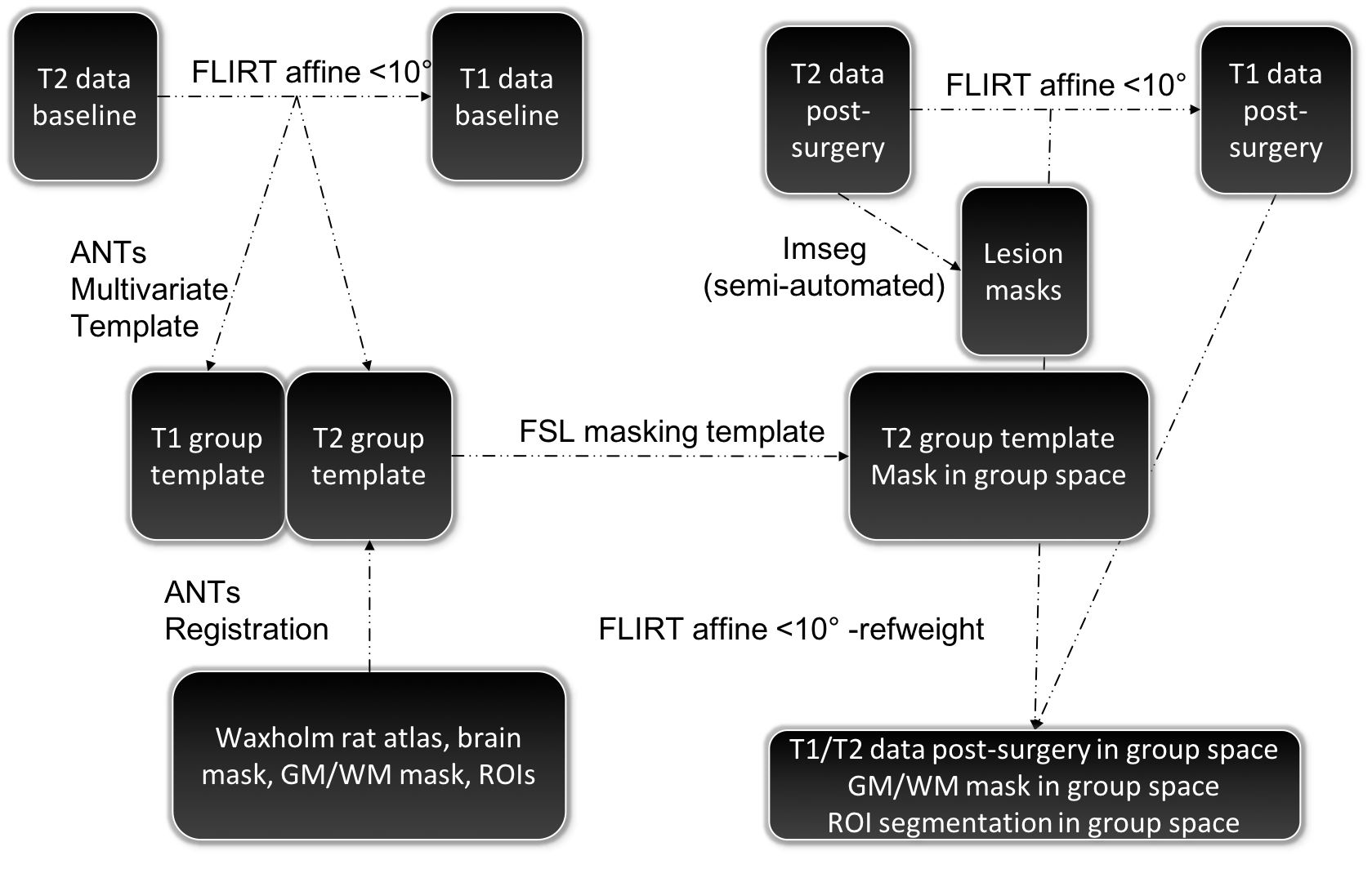
Registration Baseline-timepoint images: T2 baseline images were registered to their T1 counterparts (FSL-FLIRT affine-only registration, limiting the angle rotations to 10° or less). A group template was then created using T1 and T2 images in T1 space with ANTs (antsMultivariateTemplateConstruction.sh). The publicly available rat atlas3 (Waxholm Space Atlas, containing T2/DWI data, brain and GM/WM masks) was registered to the T2 group template (ANTs, non-linear). Injury-timepoint images: T2 images were registered to their T1 counterparts similarly to before. Brain extraction was performed in T1 individual space using antsBrainExtraction.sh (with the previously created T2 group template and mask as inputs). Semi-automatic segmentation, using IMSEG v1.8, was conducted to delineate brain areas with focal lesions in the T2 individual space images. Finally, an affine-only registration was performed on the resulting skull stripped T2 images to the group template space using FSL-FLIRT, with the lesion masks supplied as argument to the -inweight parameter, plus limited angles < 10°.
Statistics Voxel-wise comparisons were performed using FSL randomise (10k permutations) in the group space separately for T1 and T2 weighted contrasts. T-tests were performed to investigate an interaction between group by testing for an intensity change between baseline and post-surgery animals. Analysis was performed separately for white matter and grey matter using binarised masks from the WAXHOLM atlas.
RESULTS:
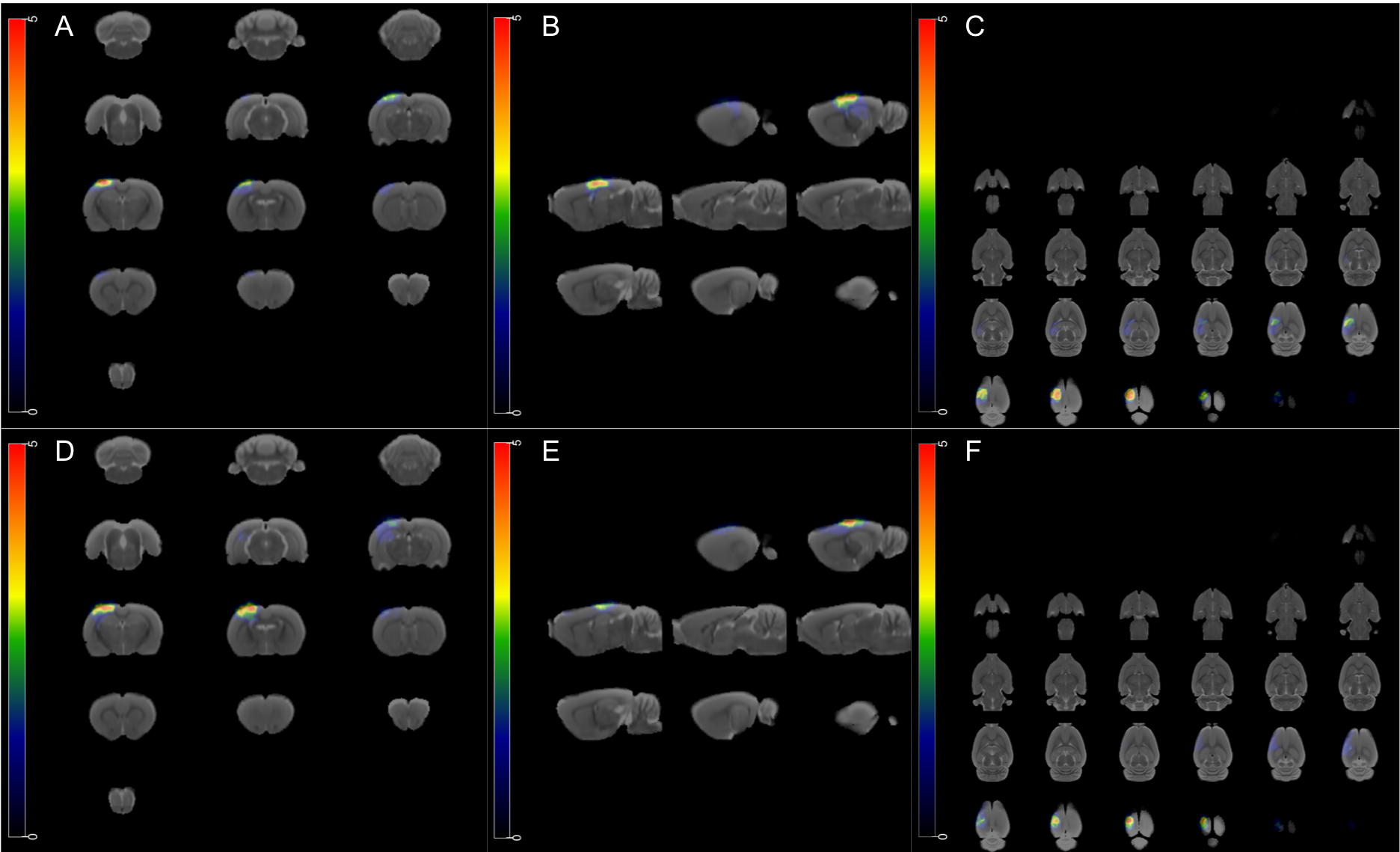
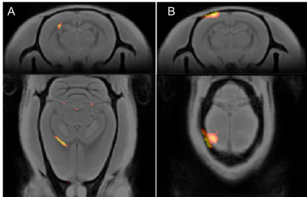
Figure 1 describes the registration pipeline, illustrating the steps from individual T1/T2 space, to baseline group template creation, to registration of the injured rat brains with lesions to the template. Figure 2 shows the position and prevalence of lesions following CCI surgery, for the mild and moderate groups separately. An increased number of animals displayed visible bigger injuries in T2-weighted images in the moderate group (2mm injury depth), but the individual lesions were variable. Figure 3 illustrates preliminary voxel-wise group comparisons between baseline and injury. No significant differences were found in T1. T2 showed significant differences in the grey matter periphery of the impact and in the ipsilateral corpus callosum white matter.DISCUSSION:
The CCI surgery caused skull deformations in several cases, which made brain extraction necessary to achieve registration. Automated brain extraction is one of the most challenging steps with rodent brains, with common human methods (ie FSL BET), not applicable. Other methods (freesurfer/Ants) require an initial segmentation or template to be successful. In this pipeline, the group template was created using a combination of the T1 and T2 modalities, an optimised affine registration and an existing atlas. T2 had a higher contrast between tissue borders, but adding T1 when building the group template improved the result. Preliminary voxel-wise comparisons showed significant grey matter alterations in the area of impact, reflecting the edema visible in T2-weighted images, whilst the white matter changes showed damage in the ipsilateral corpus callosum tract. Future analysis will include ROI-based volume changes estimations (SPM), based on atlas-defined regions.CONCLUSION:
Our results illustrate a robust pipeline for registration of challenging preclinical rat brains with pathological lesions, using established MRI software including FSL and ANTs. Alternatives to semi-automatic segmentation of the lesions in the T2 images would be desired as the next step for a fully automated analysis pipeline.Acknowledgements
No acknowledgement found.References
1. Dixon CE, Clifton GL, Lighthall JW et al. A controlled cortical impact model of traumatic brain injury in the rat. J Neurosci Methods. 1991;39(3):253‐62.
2. Crum WR, Modo M, Vernon AC et al. Registration of challenging pre-clinical images. Computational Neurosci. 2013; 216(1):62-77.
3. Papp EA, Leergaard TB, Calabrese E et al. Waxholm Space atlas of the Sprague Dawley rat brain. NeuroImage. 2014; 97:374-386.
Figures