3216
Ultra-High-Resolution Diffusion Tensor MRI Detects Early Axonal Connectivity Anomalies in Hippocampal Regions of ALS Mice1Bioengineering, University of Illinois at Chicago, Chicago, IL, United States, 2Anatomy and Cell Biology, University of Illinois at Chicago, Chicago, IL, United States, 3Department of Physics, University of Florida, Gainesville, FL, United States, 4Department of Pathology, Northwestern University, Chicago, IL, United States, 5Department of Biochemistry and Molecular Biology, University of Florida, Gainesville, FL, United States
Synopsis
Ultra-High field (UHF) MRI has been in continuous development as a new tool to investigate ultrastructural microscopic details in neuropathology. In this study we use UHF-MRI (17.6T) to investigate presymptomatic changes in the hippocampus of animal models of ALS (G93A-SOD1 mice). Using an ALS fluorescent transgenic mouse reporter and optical confocal microscopy, we demonstrated that microstructural changes detected by MRI diffusion can be related to very early alterations in axonal connectivity. This study constitutes a stepping stone for the application of more complex diffusion models in inhomogeneous brain tissue as a non-invasive exploration of neuropathology in ALS.
TARGET AUDIENCE
Neuro-radiologists with expertise in MRI diffusion applied to neurodegenerative disorders.INTRODUCTION
Amyotrophic lateral sclerosis (ALS) is a genetic disorder characterized by a progressive decline in motor functions 1. However, a portion of patients with ALS have also shown a significant decline in cognitive functions, implying a cerebral circuitry disconnection. Ultra-high field MRI (UHF-MRI) has been proven a very important imaging tool to capture the microstructural changes in deep white and grey matter regions in the context of ALS2. In this study we propose to use UHF-MRI as a tool to investigate early (presymptomatic) microstructural changes in an animal models of ALS (G93A-SOD1 mice). We hypothesize that microstructural changes observed in UHF-MRI can be related to alterations in axonal connectivity visualized using microscopy techniques.
METHODS
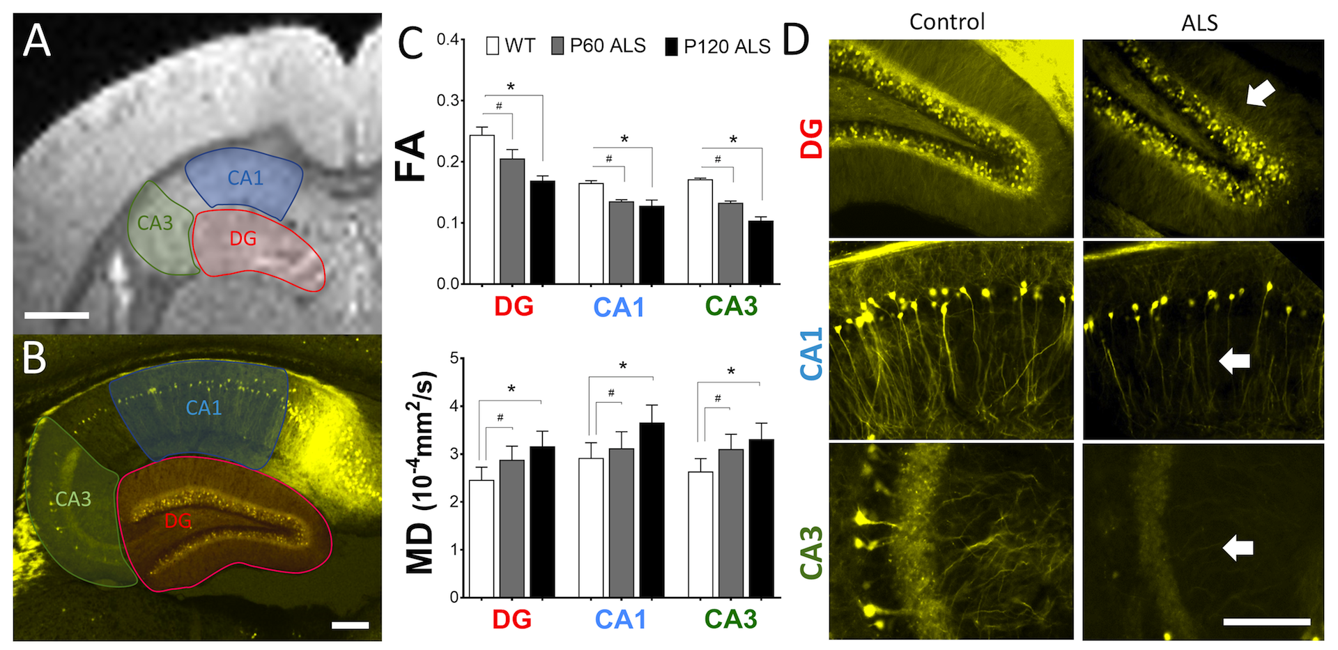
Specimen Preparation: Mice brains were obtained at 60 and 120 days of age (P60 presymptomatic and P120 symptomatic groups) in accordance with institutional animal care and committee regulations. MRI studies: Paraformaldehyde-fixed brains from transgenic yellow fluorescent (YFP) control mice (n = 4) and YFP, G93A-SOD1 mice (ALS mice) from P60 and P120 mice (n = 4 per group) were washed from PBS and immersed in Fluorinert TM oil (#FC43, 3MTM). Three mice brains were vertically placed inside 10 mm NMR tubes. To minimize variability between scans and cost, we included three 10 mm NMR tubes (New Era, NJ) inside a 25mm RF coil (450 mT/m) per scanning session (total = 2). Image Acquisition: MRI scanning were achieved using a 17.6 T, 89 cm bore Bruker Avance III HD 750 MHz MRI scanner with a 25 mm Quad Transceiver mouse coil. Careful manual shimming was performed before diffusion measurement. Such measurements were obtained by using a spin-echo diffusion weighted sequence with the following acquisition parameters: TR = 10,000 ms, TE = 20 ms, , FOV = 25x25x34 mm, matrix size = 125x125x170, image resolution 150x150 μm and b = 1000, 2500 s/mm2, δ/Δ = 3.5/11 ms), averages = 3. We used 20 and 64 directions of diffusion gradients, DWI acquisition time was 6 hours for b = 1000 and 19 hours for b = 2500. Histological Analysis: Brains were processed for confocal fluorescence microscopy according to methods previously described 3. Data Analysis: Fractional anisotropy (FA) and mean diffusivity (MD) values were extracted from the experimental data FSL software package. These values were computed from the ROIs drawn on the slice. Imaging analysis by FSL algorithms (Analysis Group, FMRIB, University of Oxford, UK) was used to generate FA and MD outputs and ImageJ software (NIH, Maryland, USA) to manually segment each hippocampus ROI. Statistical analysis using ANOVA and post-hoc Tukey test (GraphPad Prism 6.0 software).
RESULTS
A decrease in FA was observed in the dental gyrus (DG) portion of the hippocampus between control mice (YFP) and ALS mice (YFP, G93A-SOD1) at early stages of the disease (P60) (YFP mice = 0.24+/-0.01 vs. ALS mice = 0.19+/-0.05) (p<0.05) (-21%). An additional decrease in FA was also noted in ROIs located in the regions of Cornu Ammonis-1 (CA1) and Cornu Ammonis-3 (CA3) of the hippocampus, (YFP mice = 0.16+/-0.02 vs. ALS mice = 0.13+/-0.04) (p<0.05) (-18%) and (YFP mice = 0.17+/-0.01 vs. ALS mice = 0.10+/-0.02) (p<0.05) (- 40%). A further decrease in FA was also noted between YFP mice and symptomatic ALS mice (P120). In addition, DTI studies also evidenced an increase in MD between control and ALS mice not only in the DG region (YFP mice = 2.9+/-0.05 x10-4 vs. ALS mice = 3.4+/-0.02 x10-4) (p<0.05) (+17%), but also in the CA1 region (YFP mice = 3.4+/-0.06 x10-4 vs. ALS mice = 3.8+/-0.04 x10-4) (p<0.05) (+12 %) and CA3 region (YFP mice = 2.5+/-0.03 10-4 vs. ALS mice = 3.9+/-0.04 10-4) (p<0.05) (+56%) respectively. Confocal fluorescence histological analysis in the YFP, G93A-SOD1 also demonstrates anomalies in dendrite and axonal morphology in all interrogated regions (Fig.1).
DISCUSSION
Previous MRI diffusion studies have shown that changes in diffusion tensor imaging (DTI) parameters are related to central nervous system alteration in gray and white matter regions of ALS mice3. To the best of our knowledge, this is the first study combining UHF-MRI and confocal microscopy to study axonal connectivity in the hippocampus of ALS mice. Our results showed that microstructural changes detected by UHF-MRI relate to early anomalies in neuronal connectivity and that MRI diffusion constitutes a useful imaging technique for the early detection of histological changes in ALS. This study constitutes the initial stage for the applications of more complex diffusion models to interrogate ALS neuropathology in the brain.
Acknowledgements
This study was supported in part by a Chicago Biomedical Consortium (CBC) postdoctoral fellowship grant (Award #085740) to RG at the University of Illinois in Chicago. Data collection was supported by the National Science Foundation Cooperative Agreement No. DMR-1157490 and the State of Florida, Magnetic Laboratory Visiting Scientist Program (Award VSP #278) to RG. A portion of this work was performed in the McKnight Brain Institute at the National High Magnetic Field Laboratory (NHMFL) and Advanced Magnetic Resonance Imaging and Spectroscopy (AMRIS). Animal husbandry was done at the Department of Anatomy and Cell Biology, University of Illinois at Chicago. Histological studies were realized at the Department of Pathology, Feinberg School of Medicine, Northwestern University.
References
1. Gatto RG, Li W, Magin RL. Brain Res. 2018:(1679)45-5. 2. Gatto RG, Amin MY et al. Transl. Neurodegener. 2018:(7)20. 3. Gatto RG, Li W, Gao J, Magin RL et al. NMR Biomed. 2018:(8): e3954.
Figures