3215
In Vivo MRI characterization of the effect of neuroprotection after nerve agent poisoning in rats1Radiology & Radiological Sciences, Uniformed Service University, Bethesda, MD, United States, 2Center for Neuroscience and Regenerative Medicine, Henry M. Jackson Foundation, Bethesda, MD, United States, 3Anatomy, Physiology and Genetics, Uniformed Service University, Bethesda, MD, United States, 4Center for Neuroscience and Regenerative Medicine, Uniformed Service University, Bethesda, MD, United States
Synopsis
Organophosphate poisoning is a major public health problem in developing countries. Organophosphates cause prolonged seizures and leads to neurodegeneration and functional defects. Current therapies are largely ineffective. Several advanced neuroimaging techniques demonstrate promise to detect subtle changes in brain activity and morphology related to organophosphate nerve agent poisoning, which would allow for the in vivo assessment of new therapeutics. In this study we use in vivo MRI and immunohistochemistry to demonstrate that fluorinated volatile anesthetics are an effective post-exposure neuroprotectant and can be used for organophosphates poisoning treatment.
Introduction
Organophosphate (OP) poisoning results in significant morbidity and mortality. OP agents are categorized as potential weapons of mass destruction1. OP poisons elevate acetylcholine at neuromuscular junctions throughout the central nervous system (CNS), resulting in prolonged seizures and neuronal loss. After 20 minutes this cholinergic phase transitions into a pathophysiology dominated by glutamatergic overactivation2, and results in substantial neurodegeneration and permanent CNS damage. Currently there are no therapies which are satisfactorily effective in the later, non-cholinergic phase of OP poisoning3.
Fluorinated volatile anesthetics have a wide array of actions in the CNS, including neuroprotective and anti-inflammatory effects. Recently, we found that the problem of glutamatergic overactivation can be reduced effectively using fluorinated volatile anesthetics such as isoflurane4. Therefore, isoflurane represents an excellent candidate drug for the treatment of the CNS pathologies resulting from OP poisoning.
Magnetic Resonance Imaging (MRI) is capable non-invasively revealing key pathological processes in animals exposed to a nerve agent. This allows us to characterize nerve agent-induced brain damage and to assess the efficacy of new therapies in vivo.
The goal of this study was to use in vivo neuroimaging and immunohistochemistry to ascertain if volatile anesthetics protect the CNS from OP-induced damage when administered 1-hour post toxin exposure at anesthetic doses. This makes this treatment immediate applicable and available in hospitals and on the battlefield.
Methods
Male Sprague-Dawley rats (n=41) were randomly assigned to control, non-treated, and isoflurane treated groups. Paraoxon (POX) was administered (S/C) with atropine and (Pralidoxime (2-pyridine aldoxime methyl chloride) 2-PAM (IM). The treated group received 5% isoflurane for 5 min at 1-hour post POX exposure. Rats were continuously rated for seizure severity using modified Racine Scale.
MRI experiments were conducted 24 hours after POX exposure and evaluated for brain hyper-intense parenchymal lesions, parenchymal edema, and BBB integrity using a 7T Bruker Biospec 70/20 (Bruker Biospin, Billerica, MA).
The 2D multi-echo Rapid Acquisition with Relaxation Enhancement (2D RARE) sequence with repetition time TR=5500 ms, echo time TE = 10, 30, 50, 70, 90, 110 ms, echo train length ETL=2, in-plane 150x150 µm2, slice thickness Thk=750 µm, was used for T2 map computation to assess vasogenic edema. DTI indexes were computed based on 2D single-shot echo planar imaging acquisition, TR=8000 ms, TE=32 ms, 15 diffusion directions, b=1000 s/mm2, Δ/δ =12/4 ms , in-plane 300x300 µm2, Thk=750 µm to evaluate cytotoxic edema. 2D RARE with TR=1400 ms, TE=5 ms, ETL=4, inversion time TI=1400 ms, in-plane 150x150 µm2, Thk=750 µm was used for acquiring T1W pre and post contrast agent (CA) IP injection (0.5 ml, ProHance 0.4 mmol/kg) to appraise the integrity of the blood brain barrier (BBB).
MRI findings were correlated with histology using GFAP, caspase-3 markers and Fluoro Jade B (FJB). Two-way ANOVA statistical tests (Sidak's) were used for analysis.
Results and Discussion
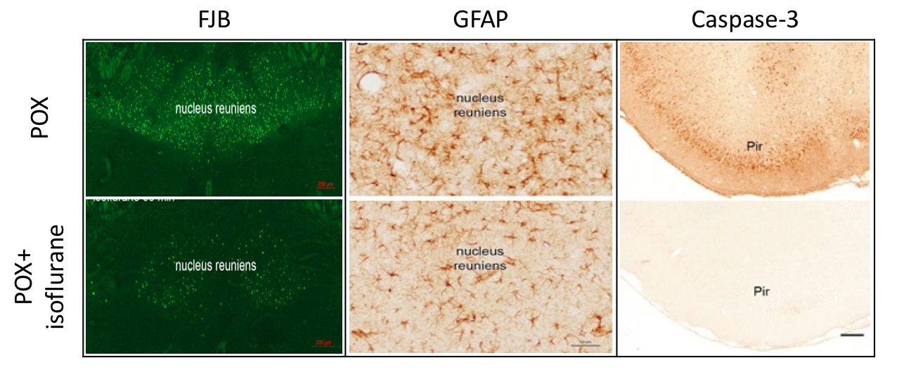
FJB staining, as well as GFAP staining, was extensive in several brain areas of surviving untreated animals 24 hours after POX administration indicating neuronal degeneration and a shift in astrocyte morphology to that of reactive astrocytes. In contrast, isoflurane treated animals showed greatly reduced FJB and GFAP staining’s with the astrocytes exhibiting resting morphology. Caspase-3 staining was low or absent in control animals and a substantial reduction in caspase-3 was observed in isoflurane treated animals (Figure 1).
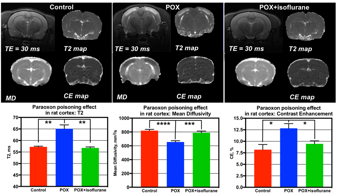
T2-weighted hyperintense lesions co-located with diffusion-weighted hyper-intense lesions (indicative of cytotoxic edema) in neocortex, piriform cortex and hippocampus were apparent in untreated poisoned animals. The isoflurane treated animals had minimal brain parenchymal hyper-intense lesions (Figure 2). Cortical T2 and mean diffusivity values derived in POX+Isoflurane group were similar to controls. The reduced contrast enhancement (CE) in isoflurane treated animals is an indication of BBB protection by isoflurane after POX poisoning.
The lesions shown by MRI were consistent with the lesions seen with FJB, caspase-3 and GFAP immunostaining. Extensive neuronal degeneration occurred in the untreated animals 24 hours after POX administration, whereas isoflurane treated animals had minimal brain lesions, comparable to controls, which was confirmed by MRI and histopathology staining’s.
Summary
In vivo MRI derived T2, mean diffusivity, and CE maps were able to distinguish subtle changes in the brain which was highly correlated with the histopathological findings, making them a valuable tool in diagnosing the neurodegenerative phase of organophosphate poisoning. This study demonstrates that fluorinated volatile anesthetics are neuroprotective in OP poisoning and capable of halting OP poisoning-induced seizures and resultant neurodegeneration. Broad availability of volatile anesthetics and the non-invasive nature of our approach makes it attractive for field applications in military operations, accidental exposure or terrorism.Acknowledgements
This work was funded by the U.S. DOD in the Center for Neuroscience and Regenerative Medicine and NIH grant NS 076448.References
1. Jett DA (2007): Neurological aspects of chemical terrorism. Ann Neurol 61: 9-13.
2. Torres-Altoro MI, Mathur BN, Drerup JM, Thomas R, Lovinger DM, O'Callaghan JP, et al (2011): Organophosphates dysregulate dopamine signaling, glutamatergic neurotransmission, and induce neuronal injury markers in striatum. J Neurochem 119: 303-313.
3. Buckley NA, Eddleston M, Li Y, Bevan M, Robertson J (2011): Oximes for acute organophosphate pesticide poisoning. Cochrane Database Syst Rev : CD005085.
4. Krishnan JKS, Figueiredo TH, Moffett JR, Arun P, Appu AP, Puthillathu N, et al (2017): Brief isoflurane administration as a post-exposure treatment for organophosphate poisoning. Neurotoxicology 63: 84-89.
Figures