3214
Spectroscopy and sodium analysis of dissociated cellular therapy in acute ischemia at 21.1 T1National High Magnetic Field Laboratory, Florida State University, Tallahassee, FL, United States, 2Chemical & Biomedical Engineering, FAMU-FSU College of Engineering, Tallahassee, FL, United States
Synopsis
This study evaluates biochemical markers in a rat model of acute ischemia at 21.1 T following administration of human mesenchymal stem cells dissociated (d-hMSC) from 3D aggregates utilizing sodium (23Na) chemical shift imaging (CSI) and relaxation-enhanced MRS. High field 1H MRS provides sensitive longitudinal metabolic mapping of biological markers including lactate, NAA, creatine and choline in response to cellular treatment. Evaluation of 23Na CSI provides insight into cerebral ionic homeostasis and tissue recovery following acute neurodegeneration by tracking ischemic lesion volumetrics.
Introduction
The ability to assess treatment longitudinally and establish early indicators for tissue recovery is imperative to evaluating the efficacy of novel therapies for stroke.1 Here, human mesenchymal stem cells dissociated from 3D aggregates (d-hMSC) are injected intra-arterially (IA) in a rat model of transient ischemia to identify biochemical markers by MRS and sodium (23Na) chemical shift imaging (CSI). Applying these techniques at ultra-high fields to evaluate stroke recovery provides, with increased sensitivity, insight into ionic and metabolic homeostasis indicative of tissue recovery.2 Furthermore, ultra-high field spectroscopy utilizing a relaxation-enhanced (RE) MRS method for selective excitation provides a highly sensitive approach to monitor metabolic concentrations and provide a potential early marker for tissue recovery.3 We hypothesize that administration of d-hMSC will enhance recovery and provide restoration of ionic and metabolic homeostasis as indicated by 23Na CSI and RE-MRS.Methods
Cell Source: hMSC were acquired from the Tulane Center for Stem Cell Research and Regenerative Medicine. A range of aggregate sizes approximately 400-µm in diameter was systemically induced in ultra-low attachment well plates under rocking motion for 2 days. Aggregates then were dissociated 48 h prior to implantation and exposed to 7.47 µg Fe/mL for labeling with micron-sized particles of iron oxide for 12 h immediately prior to administration.
Animal Model: Transient Middle Cerebral Artery Occlusion (MCAO) was instituted for 1 h in Sprague-Dawley rats (200-250 g) to induce ischemia.4 Following MCAO, the animals were either administered 1 million d-hMSC suspended in phosphate saline buffer (PBS) (d-hMSC+MCAO; n=4) or PBS only (PBS+MCAO; n=2) via microneedle injection through the MCA.
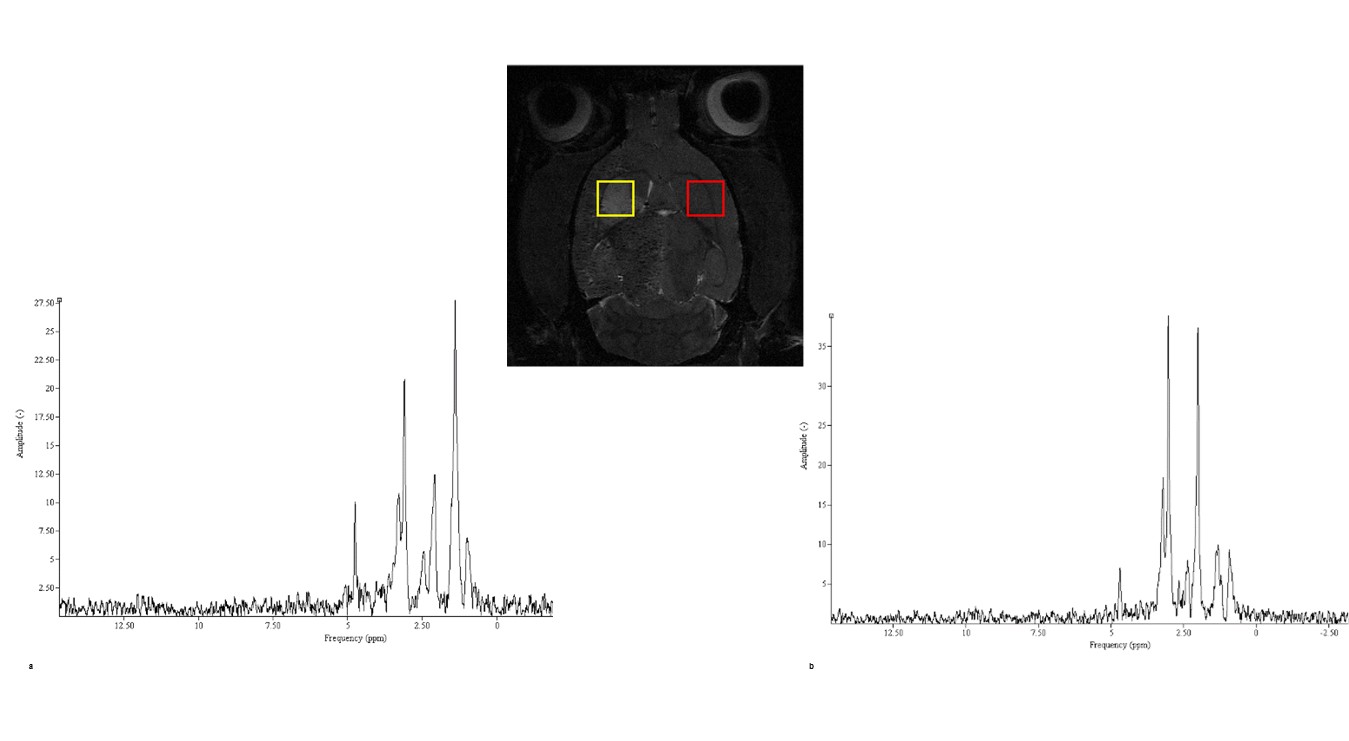
MR Acquisitions: High resolution MR imaging and spectroscopy were acquired in vivo on day 1, 3 and 7 post-MCAO utilizing the 21.1-T magnet at US National High Magnetic Field Laboratory (NHMFL). 3D 23Na CSI was acquired at 1-mm isotropic resolution with TR = 60 ms. RE-MRS, which utilizes a selective spectral excitation approach3 was used to evaluate metabolites in a 3-mm isotropic voxel in both ischemic and contralateral hemispheres. A 4-kHz bandwidth excitation pulse was used to target metabolites of interest (Lac, Cre, Cho and NAA) while avoiding water. Localization by an adiabatic selective refocusing (LASER)3 pulse sequence was utilized for spatial selectivity. Anatomical reference to the ischemic core and contralateral alignment was achieved by referencing T2-weighted images generated from a 1H Fast Spin Echo (FSE) sequence with 100x100-µm in plane resolution (Fig.1).
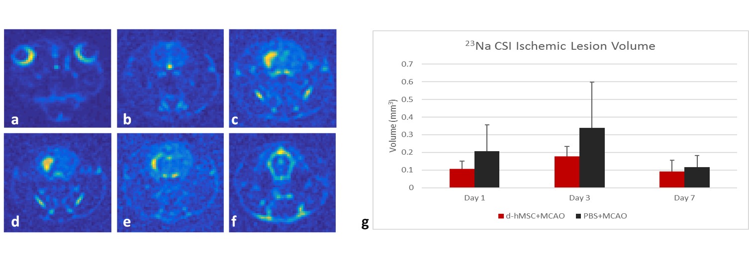
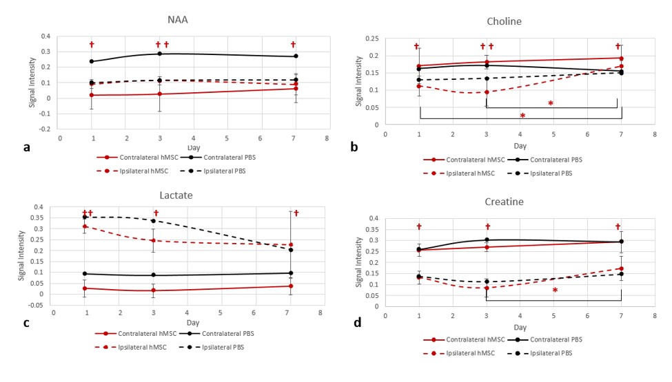
Analysis: CSI data reconstructed in MATLAB was zero-filled to 0.5-mm isotropic resolution as shown in Fig.2. Lesion volumes were quantified based on contralateral 23Na signal and standard deviation (SD) obtained from day 1 according to the following: Signalischemia = Signalcontralateral + 2.5SD. RE-MRS data was reconstructed in JMRUI using the Linear Prediction Singular Value Decomposition algorithm to select components within the zero-filled (16k) and line broadened spectra (Fig.1). Peaks were assigned according to previous literature for metabolites of interest (NAA 2.0 ppm, Lac 1.31 ppm, Cre 3.0 ppm, Cho 3.2 ppm)5 referencing water at 4.7 ppm. Absolute signal intensities were used to track metabolite concentration longitudinally between hemispheres and groups. Statistical analysis was preformed for d-hMSC+MCAO (n=4) temporally and hemispherically.
Results
23Na signal and lesion reduction on day 7 (Fig.2) demonstrate potential tissue recovery and return of sodium homeostasis. Contralateral signal intensities indicate no significant change temporally for all metabolites in the d-hMSC treated group, indicating maintenance of baseline metabolites. For d-hMSC+MCAO, Cho and Cre demonstrate reductions ipsilaterally compared to the contralateral hemisphere, with noticeable trends towards equilibrium on day 7 (Fig.3). Lac and NAA significantly increased immediately following ischemia with reduction of differences for both metabolites at day 7.Discussion
Preliminary 23Na CSI data indicate volume reductions with additional repetitions underway to determine statistical significance. The initial spike in ipsilateral Lac is reduced at subsequent time points but appears to return toward the contralateral level more rapidly for d-hMSC+MCAO, indicative of energetic recovery in the lesion (Fig.3c). Following an initial decrease in both Cre (Fig.3b) and Cho (Fig.3d), metabolite recovery was demonstrated both longitudinally and between hemispheres. PBS-MCAO demonstrated elevated contralateral Lac signal compared to d-hMSC-MCAO, potentially indicative of global ischemia effects that are attenuated by d-hMSC.Conclusion
23Na lesion volume decreases following an initial spike after ischemia has been shown to be a sensitive parameter for tissue recovery evaluation. In combination with metabolic assessment via RE-MRS, we aim to provide a sensitive evaluation of early biomarkers to be established for assessing novel therapies. Preliminary results indicate d-hMSC administered animals return to homeostasis on day 7 for NAA and choline. With increased sensitivity at 21.1 T, additional metabolites will be evaluated.Acknowledgements
All work has been conducted in accordance with the FSU Animal Care and Use Committee. This work was supported by the NIH (RO1-NS102395). The National High Magnetic Field Laboratory is funded by the NSF (DMR-1644779) and the State of Florida.References
1. Benjamin EJ, et al. 2017. AMA, Statistics Committee and Stroke Statistics Subcommittee.
2. Lee V., et al. 2012. Translational Stroke Research. 3(2):236-245.
3. Shemesh N., et al. 2014. Nature Communications. DOI: 10.1038/ncomms5958.
4. Longa E.Z., et al. 1989. Stroke. 20(1):84-91.
5. Govindaraju V., et al. 2000. NMR in Biomed. 13:129-153.
Figures