3213
Serial MRI to Assess Effects of Drug Particle Size on Inflammation and Pharmacokinetics to Support Development of Long Acting Parenteral Formulations1Bioimaging, GlaxoSmithKline, Collegeville, PA, United States, 2Histology, GlaxoSmithKline, Collegeville, PA, United States, 3Pathology, GlaxoSmithKline, Collegeville, PA, United States, 4Modeling and Translational Biology, GlaxoSmithKline, Collegeville, PA, United States, 5Drug Delivery, GlaxoSmithKline, Collegeville, PA, United States
Synopsis
We evaluated the effect of Long Acting Parenteral particle size on drug depot kinetics and inflammation using ultra small paramagnetic iron oxide (USPIO) T2W MRI. Our results showed an immediate post injection difference in inflammatory response and histological confirmation of greater muscle injury in the smaller micronized (1um) particle group compared to larger 20um particle formulation. The imaging of the drug depot in vivo with MRI combined with drug PK, tissue biodistribution, and histology allows for the development of individual Physiological Based PK models of drug biodistribution which would add significant scientific value to drug development.
Introduction
Advances in solid drug nanoparticle technologies have resulted in a number of long acting parenteral (LAP) formulations for once monthly or longer administration to treat schizophrenia1, opioid addicition2, and as oral contraceptives3. LAP formulations offer great utility and benefit for chronic diseases, especially when a lack of medication compliance may be detrimental to the treatment response4. In order to understand how inflammation following LAP intramuscular injection may affect compound pharmacokinetics (PK), we evaluated the effect of particle size on drug depot kinetics and inflammation using ultra small paramagnetic iron oxide (USPIO) T2W MRI to support developing LAP formulations.Methods
Three groups of male Sprague Dawley rats were given GSK2838232 by IM injection in one of two particle sizes (Micronized (20 um) or Micronized (1 um), or Micronized (1 um) + intravenous USPIO, (n=4/group) into the right gastrocnemius muscle at Day 0). Vehicle control (0.2% Tween-80, 2% PEG3350, 4.5% Mannitol, 10 mM PBS, pH 7.2) was injected in left gastrocnemius muscle. The dose volume was 182 ul/kg and the drug concentration was 110 mg/ml. USPIO (Ferumoxytol) dose was 1000 umol/kg used to assess the macrophage uptake in the depot. A 4.7T magnet (Bruker Biospin) with a 72mm resonator coil was used to acquire serial T2W images to assess depot volume for right and left injection sites out to 15 days post IM injection. A RARE sequence with fat suppression with scan parameters (TR/TE=2000/20ms (effective TE=60ms), RARE factor=8, bandwidth=50kHz, FOV=10cmx10cm, matrix=128, slice thickness=2mm, NEX=8, total acquisition time=4min 16 secs was used in this study. The study design for the time points for serial blood PK sampling, imaging, and terminal histological evaluation of injection sites are shown in Fig. 1. All studies were conducted in accordance with the GSK Policy on the Care, Welfare and Treatment of Laboratory Animals and were reviewed by the Institutional Animal Care and Use Committee at GSK.Results
At the 24 hour post injection timepoint the drug depot volumes were significantly larger in the 1 um groups compared to 20 um group, whereas there was no depot in the vehicle control group. The drug depot volumes remained significantly larger at Day 15 (Fig. 2). At the 24 hour timepoint post USPIO injection depot signal intensity reflects vascular leakage in the depot which slowly changes over 12 days as macrophage associated iron signal intensity is reflected around the perimeter of the depot (Fig. 3). Histological analysis of injection sites revealed less severe muscle injury, evidenced by amount of necrosis and inflammation with encapsulation, in the 20 um group compared to the 1 um group (Fig. 4). Blood PK was similar between 20 um and 1 um groups. Both groups remained above the IC90 for the 15 day study (Fig. 5).Conclusions
The 20 um particle size induced significantly smaller depot volumes at Day 1 (~ 1/3 the depot volume) compared to 1 um groups and this difference was maintained over the 2 week study. This result was consistent with a reduced severity of muscle injury at the injection site in the 20 um group, reflected by the severity of necrosis and inflammation. However, there were no adverse clinical signs in any treatment group. Macrophage accumulation around the perimeter of the depot was identified in the USPIO group. Blood PK profiles (AUC) were similar between groups (Both groups remained above the IC90 for the 15 day study). The larger micronized particle formulation produced less muscle injury and similar PK profiles compared to the smaller micronized formulation, indicating no advantage to the smaller micronized formulation to increase systemic exposure of the drug in this study. The imaging of the drug depot in vivo with MRI combined with drug PK, tissue biodistribution, and histology allow for the development of individual Physiological Based PK models of drug biodistribution which would add significant scientific value to drug development.
Acknowledgements
No acknowledgement found.References
1Suzuki T. Exp Opin on Drug Delivery 13 (2016) 253-64,2Wu L et al.Pharm Research 32(2015) 2180-91, 3Gastfriend D.Ann N.Y.Acad.Sci. 1216 (2011) 144-66, 4Owen A. et al. Adv Drug Delivery Reviews 103 (2016) 144-56.
Figures
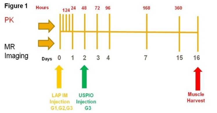
Figure 1-Study Design-Time points for serial blood PK sampling, imaging, and terminal histological evaluation of injection sites.
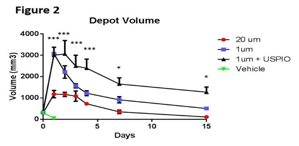
Figure 2- MRI Depot Volume- 24 hours post injection, depot volume was significantly larger in the 1 um groups compared to 20 um group; whereas there was no depot detected in the vehicle control group. 1 um + USPIO depot volumes remained significantly larger at Day 15.
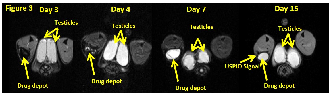
Figure 3- At 24 hours post USPIO injection (Day 3 post LAP injection), the drug depot signal intensity reflects vascular leakage in the depot which slowly changes over 12 days as macrophage associated iron signal intensity is reflected around the perimeter of the depot.
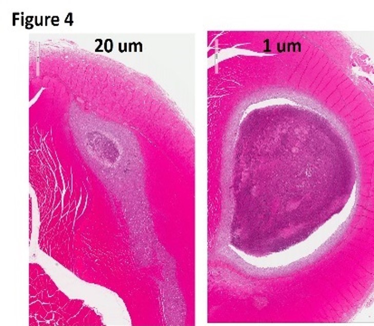
Figure 4-Histological analysis of injection sites revealed less severe muscle injury, evidenced by amount of necrosis and inflammation with encapsulation, in the 20 um group compared to the 1 um group.
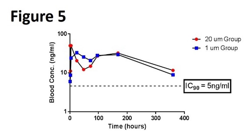
Figure 5-Blood PK was similar between 20 um and 1 um groups. Both groups remained above the IC90 for the 15 day study.