3210
Using a multimodal near-infrared spectroscopy and MRI system to quantify gray matter metabolic rate for oxygen: A hypothermia validation study1Biomedical Engineering Graduate Program, University of Calgary, Calgary, AB, Canada, 2Department of Radiology, University of Calgary, Calgary, AB, Canada, 3Hotchkiss Brain Istitute, University of Calgary, Calgary, AB, Canada, 4Experimental Imaging Centre, University of Calgary, Calgary, AB, Canada, 5Cumming School of Medicine, University of Calgary, Calgary, AB, Canada
Synopsis
Non-invasive quantitative imaging of cerebral oxygen metabolism (CMRO2) in mice is crucial to understand the role of oxidative metabolism in neurological diseases. We are developing a multimodal method combining near-infrared spectroscopy and high-field MRI to non-invasively study oxygen delivery and consumption in the cortex of mouse models of neurological disease. In this study, the feasibility of the NIRS-MRI technique to detect changes in CMRO2 in the mouse brain was assessed using a mild hypothermia, known to reduce metabolic rate. A decrease of 23% in CBF and 46% in CMRO2 was observed, which is consistent with previously published values.
Introduction
Abnormal Oxidative metabolism is likely to occur in most neurological diseases, such as Multiple Sclerosis1,2, Alzheimer's Disease3, Parkinson’s disease4, etc. These diseases usually involve hypoxia and mitochondrial dysfunction in the brain, as well as changes in cerebral blood flow (CBF) and cerebral metabolic rate for oxygen (CMRO2). It is a challenge to understand the causes and processes of neurological disorders and consequently, to search for new treatments. To this end, animal models, particularly mouse models, are often used. Moreover, non-invasive examination of oxygenation levels, dynamics of the blood flow, and mitochondrial status is crucial to further understand the role of these parameters in the brain of mouse models of neurological diseases.Objective
We are developing and validating a multimodal near-infrared spectroscopy (NIRS) and MRI technique, to study simultaneously, and non-invasively, physiological alterations in the brain of mouse models of neurological disease. A validation study is conducted to demonstrate the ability of this multimodal technique to measure the effect of pathological perturbations caused by neurological diseases, on cerebral oxidative metabolism. In this study, we examine the alterations occurring in CBF and CMRO2 in the mouse cortex under mild hypothermia (33˚C) conditions, compared to normothermia (37˚C).Methods
A 9.4T Bruker MRI with a 35 mm volume coil was used to non-invasively quantify absolute CBF with an arterial spin labeling (ASL) sequence; axial slices were acquired around the location of bregma using a CASL-HASTE sequence with the following parameters: TR/TE=3000/13.3 ms, matrix size = 128x128, FOV = 3x3 cm, slice thickness = 1 cm, 16 averages. In total, 4 perfusion images were collected per measurement: 2 controls and 2 tagged images, to correct for magnetization transfer. Following these, a T1 map was obtained in the same location as the perfusion slice using a RARE-VTR sequence where TE = 10 ms, TR=100, 500, 1000, 3000 and 7500 ms. The 4 perfusion images and T1 map are collected over a period of 12 min. CBF was calculated on a voxel-by-voxel basis5. We measured the total hemoglobin concentration (tHb), and the tissue oxygenation saturation (St) in mouse cortex using a custom-built broadband NIRS device and in-house developed processing algorithms. By measuring these parameters simultaneously with the CBF, CMRO2 can be quantified in mouse cortex, by applying the modified Fick principle6: $$$CMRO_2=k×CBF×\frac{4}{3}(S_a-S_t)×\frac{[tHb]}{CBV}$$$
Where k = 1.39 ml O2 / g Hb, is a factor describing the amount of O2 bound to Hb when completely saturated7, Sa is the arterial blood oxygen saturation, measured by the MouseOx MRI-compatible pulse oximeter on the thigh, CBV is the cerebral blood volume which is assumed to be 0.049 mL blood / mL tissue in the cerebral cortex, as measured in C57BL/6 mice8. During the experiment, the mice (n=6) were anesthetized with 2% isoflurane and ventilated with a mixture of 70% N2 and 30% O2. Rectal temperature, heart and breath rate were monitored. Perfusion and NIRS data were acquired at a core temperature of 37˚C followed by another measurement at 33˚C.
Results and Discussion
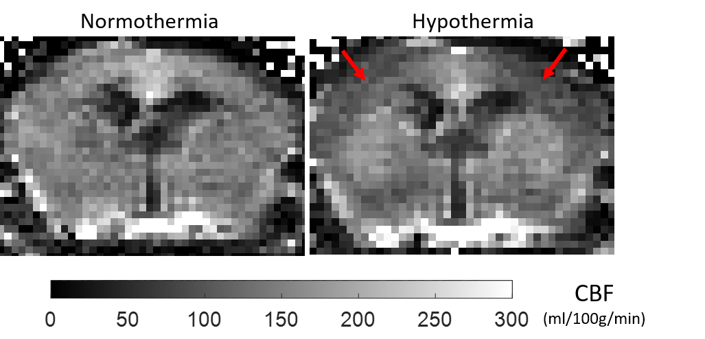
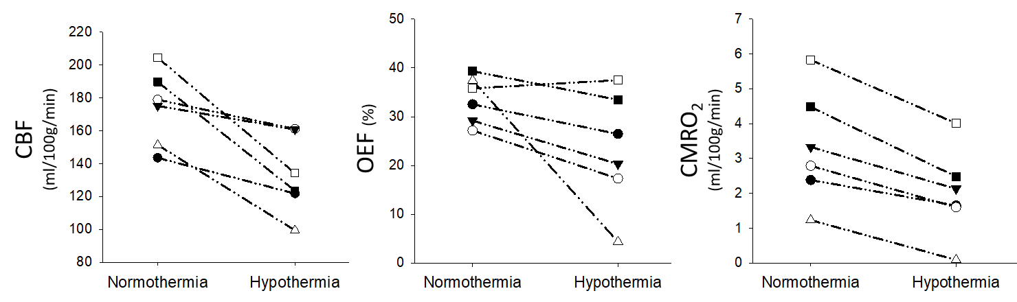
Hypothermia caused hypoperfusion in the cortex, which can be observed in the perfusion-weighted images (fig. 1). CMRO2 was 3.34±1.62 ml/100g/min at normothermia and declined significantly to 1.99±1.28 ml/100g/min (p<0.001, paired t-test) under the hypothermia condition, while CBF decreased from 174±23 ml/100g/min to 133±24 ml/100g/min (p= 0.01, paired t-test) (fig. 2). The results showed a significant decrease of 23% in CBF and 46% in CMRO2 across the cortex in mild hypothermia. This is consistent with a decrease in CMRO2 (42% ) shown previously in rats9 brains by using direct 17O NMR detection when dropping the core temperature from 37˚C to 32˚C. There was no significant change in the oxygen extraction fraction (OEF). In addition to these physiological parameters, the NIRS-MRI technique we are developing will also be capable to monitor mitochondrial status by measuring the redox state of the enzyme Cytochrome C Oxidase (CCO) in the mouse cortex.Conclusion
Here we showed that it is possible to assess absolute values of metabolic correlates such as, CMRO2, CBF, tHB, St, and OEF noninvasively in living mouse brain. This unique multimodal NIRS-MRI technique will open new possibilities for studying brain metabolism using the many mouse models of brain disease available. This method could be translated to human studies.Acknowledgements
This work is supported by the NIH R21 grant, the Natural Sciences and Engineering Research Council (NSERC), the Discovery grant, and the Biomedical Engineering Graduate program (BMEG) at U of C. Hardware are supplied by Canada Foundation for Innovation (CFI).References
1. Lin MT, Beal MF. Mitochondrial dysfunction and oxidative stress in neurodegenerative diseases. Nature. 2006;443(7113):787-795.
2. Mahad D, Ziabreva I, Lassmann H, Turnbull D. Mitochondrial defects in acute multiple sclerosis lesions. Brain : a journal of neurology. Jul 2008;131(Pt 7):1722-1735.
3. Moreira PI, Carvalho C, Zhu X, Smith MA, Perry G. Mitochondrial dysfunction is a trigger of Alzheimer's disease pathophysiology. Biochimica et Biophysica Acta (BBA)-Molecular Basis of Disease. 2010;1802(1):2-10.
4. Cai H, Cong W-n, Ji S, Rothman S, Maudsley S, Martin B. Metabolic dysfunction in Alzheimer's disease and related neurodegenerative disorders. Current Alzheimer research. 2012;9(1):5-17.
5. Buxton RB. Quantifying CBF with arterial spin labeling. Journal of magnetic resonance imaging : JMRI. Dec 2005;22(6):723-726.
6. Tichauer KM, Hadway JA, Lee T-Y, Lawrence KS. Measurement of Cerebral Oxidative Metabolism with Near-Infrared Spectroscopy: A Validation Study. Journal of Cerebral Blood Flow & Metabolism. 2006;26(5):722-730.
7. Brown DW, Hadway J, Lee T-Y. Near-infrared spectroscopy measurement of oxygen extraction fraction and cerebral metabolic rate of oxygen in newborn piglets. Pediatric research. 2003;54(6):861-867.
8. Chugh BP, Lerch JP, Yu LX, et al. Measurement of cerebral blood volume in mouse brain regions using micro-computed tomography. NeuroImage. Oct 1 2009;47(4):1312-1318.
9. Zhu X, Zhang Y, Ugurbil K, Chen W. 3D imaging of CMRO2 in rat brain at different temperature using high-field 17O NMR approach. Proceedings of International Society of Magnetic Resonance Medicine, Toronto. 2003;569.
Figures