3205
Performance of sodium, ultrafast diffusion, and MPIO stem cell tracking MRI classification of sub-acute ischemic stroke recovery at 21.1T1Medical Physics, Memorial Sloan Kettering Cancer Center, New York, NY, United States, 2Chemical and Biological Physics, Weizmann Institute of Science, Rehovot, Israel, 3Radiology, Stony Brook Medicine, Stony Brook, NY, United States, 4National High Magnetic Field Laboratory, Florida State University, Tallahassee, FL, United States, 5Chemical and Biomedical Engineering, FAMU-FSU College of Engineering, Tallahassee, FL, United States
Synopsis
MRI leverages multiple modes of contrast to characterize stroke. Acute phase stroke detection has focused on multiparametric MRI contrasts such as T2-weighting (T2w), apparent diffusion coefficient (ADC), and sodium level. Evaluation of these contrasts with theranostic cell tracking during the subacute recovery phase at ultrahigh field has not been investigated to a similar extent. Here multiparametric MRI evaluation of ADC, 23Na, and MPIO stem cell tracking in a rodent MCAO model at 21.1T was used to determine parametric correlations and receiver operator performance. Differential parametric time-dependence and sensitivities are observed that inform future high-and low field studies of stroke recovery.
Introduction
MRI leverages multiple modes of contrast to characterize stroke. Acute phase stroke detection has focused on multiparametric MRI contrasts such T2-weighting (T2w), apparent diffusion coefficient (ADC), and sodium level that exhibit differential time-dependence and sensitivities for stroke onset1. Combinations of theranostic cell tracking, ADC and sodium evolution during subacute ischemic stroke recovery have not been investigated to a similar extent. We have demonstrated that sodium and stem cell tracking is achievable at ultrahigh field2-4, and developed single-scan ultrahigh field diffusion weighted imaging approaches utilizing spatiotemporal encoding (SPEN) of the ADC5. Here we performed a multiparametric study of MCAO biomarker evolution focusing on MRI biomarker performance for stroke assessment during subacute recovery at 21.1T.Methods
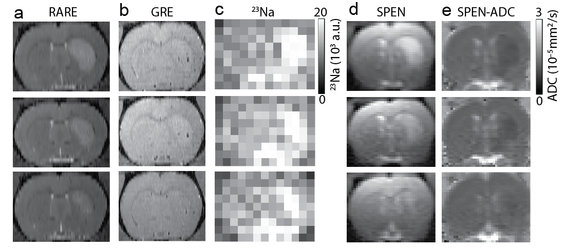
An ultra-wide bore 21.1 T (900 MHz) vertical magnet equipped with a Bruker Avance III console, Paravision 5.1 software (Bruker-Biospin, Billerica, MA) was used for all acquisitions. The system is equipped with 64-mm inner diameter imaging gradients (0.6 T/m, Resonance Research Inc., Billerca, MA) and a 33-mm homebuilt 1H quadrature surface coil or 23Na/1H resonator. 2D T2-weighted rapid acquisition with relaxation enhancement (RARE) scans acquired with an acceleration factor of 4 and 300 phase encodes, TR/TE = 6s/13ms, field-of-view (FOV) of 25.6x30 mm, and resolution of 0.10x0.10x0.5 mm was used for stroke lesion volume determination. For MPIO detection a 2D gradient recalled echo (GRE) sequence with TR/TE = 725/5 ms with FOV = 25.6 x 25.6 and resolution 0.05x0.05x0.3 mm was used. Single-slice, single-scan 2D diffusion weighted SPEN (DW-SPEN) imaging, TR=12s and effective TE=40ms=swept chirp pulse duration (sweep width=10kHz), FOV = 32x32 mm and an in-plane resolution of 0.32x0.32x2 mm was used for ADC mapping. Six b-values were used (0, 200, 400, 600, 800 and 1000 s/mm2) and applied along the x, y, and then z directions individually with Δ=9.81 and δ =3.5 ms. Cartesian sampled 3D 23Na MRI acquisitions were performed with 36 averages, TE/TR = 1/50 ms, 32x32x32 mm FOV and a matrix size of 32x32x32 resulting in 1-mm isotropic resolution for mapping sodium levels.Animal Imaging and Stem Cell Preparation
Male Sprague-Dawley rats (n=5) underwent middle carotid artery occlusion (MCAO). Human bone-marrow-derived mesenchymal stem cells (hMSCs) were cultured overnight with 0.86-m diameter iron oxide particles (micron-sized particles of iron oxide, MPIO; Bangs Laboratories, Fishers, IN) and injected intra-arterially immediately after MCAO. All animals were imaged at 1, 3, and 7 days by using RARE/GRE/DW-SPEN sequences, and at 2, 4 hrs and 8 days using RARE/23Na MRI.Statistics
Mean imaging parameters were analyzed in stroke and control ROIs using ImageJ and MatLab. Linear correlations and receiver operator characteristic (ROC) performance of each of the stroke biomarkers was determined using MatLab and GraphPad Prism.Results
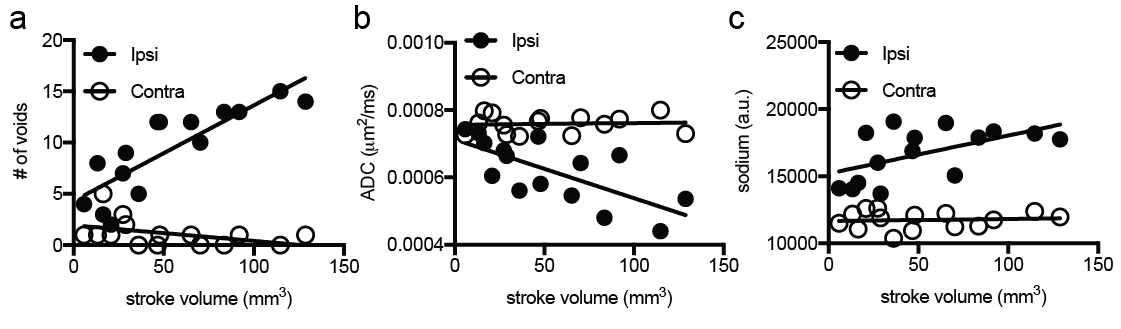
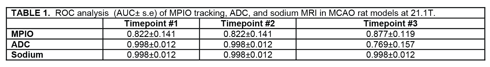
Figure 1 shows representative T2w RARE, GRE of MPIO, 23Na, SPEN-T2w, and SPEN-ADC images of the stroke at the imaging time points. These imaging parameters were analyzed over the course of the MCAO recovery and plotted as a function of stroke volume shown in Figure 2. The ROC of each of the imaging methods was calculated at each time point and areas under the ROC curves (AUC) was calculated and tabulated in Table 1.Discussion
RARE MRI was used as the benchmark for the identification of the ischemic region over the course of the study. The number of MPIO-induced voids, the ADC, and 23Na values were determined with an ROI drawn around stroke. A similar ROI was placed in the contralateral hemisphere. While each MRI parameter was associated with stroke the correlation of RARE stroke volume with these parameters varied. The ROC analysis of these measurements indicated excellent performance for stroke detection at each time point according to AUC values, but was nominally lower for the MPIO measurements, and ADC performance was reduced at 7 days post MCAO, while 23Na AUC remained unchanged over the course of the study.Conclusion
The acute phase (hours) following stroke onset is evidenced by elevated T2w contrast, reduced ADC and elevated sodium. The subacute recovery phase is characterized by reduced lesion size, increased ADC, and reduced elevated sodium area. Uptake of MPIO-labeled stem cells reports on vascular perfusion and cellular engraftment in penumbra post MCAO, while hypointense contrast at later times reflects a combination of cellular and vascular iron accumulation. Normalization of these parameters over time can reduce the sensitivity and specificity of these MRI biomarkers, but is still excellent 1-week post stroke. As MRI detection of these parameters are maximized at 21.1T, the performance of these contrasts at clinical fields and at longer recovery times can be further reduced, and may vary due to its own evolution or therapeutic intervention.Acknowledgements
This work was performed at the US National High Magnetic Field Laboratory, which is supported by National Science Foundation (Cooperative Agreements No. DMR-1157490 and DMR-1644779) and the State of Florida. Funding was also provided by the American Heart Association Grant-in-Aid 10DRNT3860040, by the Minerva Foundation Grant No. 712277 from the Federal German Ministry for Education and Research, the Kimmel Institute for Magnetic Resonance and the Perlman Family Foundation and by National Science Foundation International Research Fellowship Program, and the Fulbright Foundation.References
1. F. Wetterling, E. Chatzikonstantinou, L. Tritschler, S. Meairs, M. Fatar, L.R. Schad, S. Ansar. Investigating potentially salvageable penumbra tissue in an in vivo model of transient ischemic stroke using sodium, diffusion, and perfusion magnetic resonance imaging. BMC Neurosci 2016; 17.
2. V.D. Schepkin, W.W. Brey, P.L. Gor'kov, S.C. Grant. Initial in vivo rodent sodium and proton MR imaging at 21.1 T. Magn. Reson. Imaging 2010; 28 : 400-407.
3. J.T. Rosenberg, A. Sachi-Kocher, M.W. Davidson, S.C. Grant. Intracellular SPIO labeling of microglia: high field considerations and limitations for MR microscopy. Contrast Media Mol. Imaging 2012; 7 : 121-129.
4. J.T. Rosenberg, K.L. Sellgren, A. Sachi-Kocher, F. Calixto Bejarano, M.A. Baird, M.W. Davidson, T. Ma, S.C. Grant. Magnetic resonance contrast and biological effects of intracellular superparamagnetic iron oxides on human mesenchymal stem cells with long-term culture and hypoxic exposure. Cytotherapy 2013; 15 : 307-322.
5. A. Leftin, J.T. Rosenberg, E. Solomon, F. Calixto Bejarano, S.C. Grant, L. Frydman. Ultrafast in vivo diffusion imaging of stroke at 21.1 T by spatiotemporal encoding. Magn. Reson. Med. 2015; 73 : 1483.
Figures