3204
Upregulation of Rac1 activity related to structural and functional neuroimaging changes in mouse brain at 11.7T1Institute of Science and Technology for Brain-Inspired Intelligence, Fudan University, Shanghai, China, 2Key Laboratory of Computational Neuroscience and Brain-Inspired Intelligence, Shanghai, China, 3iHuman Institute, ShanghaiTech University, Shanghai, China
Synopsis
Rac1 is critical for synapse remodeling. In this work, we investigated whether Rac1 could induce neuroimaging changes in a transgenic mouse model using structural MRI, resting state-fMRI and MRS at 11.7T. Our data showed that the volume of the medial prefrontal cortex (mPFC) is significantly decreased in Rac1 activation group. Moreover, we found that the upregulation of Rac1 activity is related to increased functional connectivity in mouse brain. However, Glx or GABA levels do not show significant changes in mPFC. We concluded that the upregulation of Rac1 activity is related to structural and functional neuroimaging changes.
Introduction
Ras-related C3 botulinum toxin substrate 1 (Rac1) protein encoded by RAC1 gene is a signaling G protein, which belongs to the RAS superfamily of Rho family of GTPases. Rac1 functions as a pleiotropic regulator of many cellular processes, including the cell cycle, cell-cell adhesion, motility and glucose metabolism1,2. Most recently, studies have demonstrated that the activation of Rac1 is critical for synaptic structural plasticity and synapse remodeling3,4, which is closely related to brain network5. However, whether the activation of Rac1 could induce the brain structural and functional changes remains unclear. High-field MRI is a powerful in-vivo tool to noninvasively visualize anatomical and functional characteristics of rodent brains. In the present work, we aimed to investigate the association of the regulation of Rac1 activity with the changes of structural and functional neuroimaging in mouse brain using MRI at 11.7T.Materials and Methods
Animal model: All experimental protocols were approved by the Animal Ethics Committee of Fudan University. A total of 12 C57BL/6J mice (male, 8 weeks old) were obtained from Slac Laboratory (Jiaotong University, Shanghai, China). Animals were equally divided into two groups: Control (CON) group and continuous activation of Rac1 (CAR) group. To upregulate the activity of Rac1, six mice were injected with adeno-associated virus (AAV) vector with the specific shRNA for continuous activation of Rac1 (CAR) in mouse brain using microelectrodes. For the rest six mice, the empty AAV vector was injected into mouse brain as the control (CON). MRI was performed 4 weeks after AAV injection.
Data acquisition: Structural MRI, resting state-fMRI (rs-fMRI) and MRS were performed at a 11.7T BioSpec 117/16 USR MRI system equipped with a CryoProbe (Bruker BioSpin, Ettlingen, Germany). High-resolution T2W images of the whole brain were acquired using a Rapid Acquisition with Relaxation Enhancement (RARE) sequence with the following parameters: TE/TR, 30 /4400 ms; RARE factor, 8; spatial resolution, 70 × 70 × 300 μm3. The T2* data were acquired using a gradient Echo Planar Imaging (EPI) sequence with the following parameters: TE/TR, 10.5/2000 ms; spatial resolution, 200 × 200 × 300 μm3; repetitions, 240; scan time, 8 minutes. MRS was acquired using a point-resolved spectroscopy (PRESS) sequence with the following parameters: TE/TR, 12.2/1500 ms; number of averages, 512; voxel size, 1.5 × 2 × 1 mm3 located in the medial prefrontal cortex (mPFC). An isoflurane dose, ranging from 1% to 1.5 %, was used for anesthesia in all imaging runs.
Data analysis: Pre-processing was performed using SPM12 for MATLAB 2015b (MathWorks, Natick, USA). Rs-fMRI analysis were performed using home-made MATLAB code based on a previous article6, without global signal regression, band-pass filter of 0.01- 0.1 Hz. Voxel-based morphometry (VBM) was used to measure the volume of brain regions based on a mouse atlas7. MRS data were analyzed by a software, LCModel (LCModel Version 6.3; http://s-provencher.com/pages/lcmodel.shtml). Student’s t-tests were performed in MATLAB 2015b (significance, P<0.05).
Results and Discussion
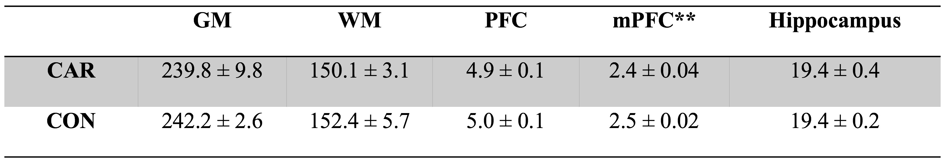
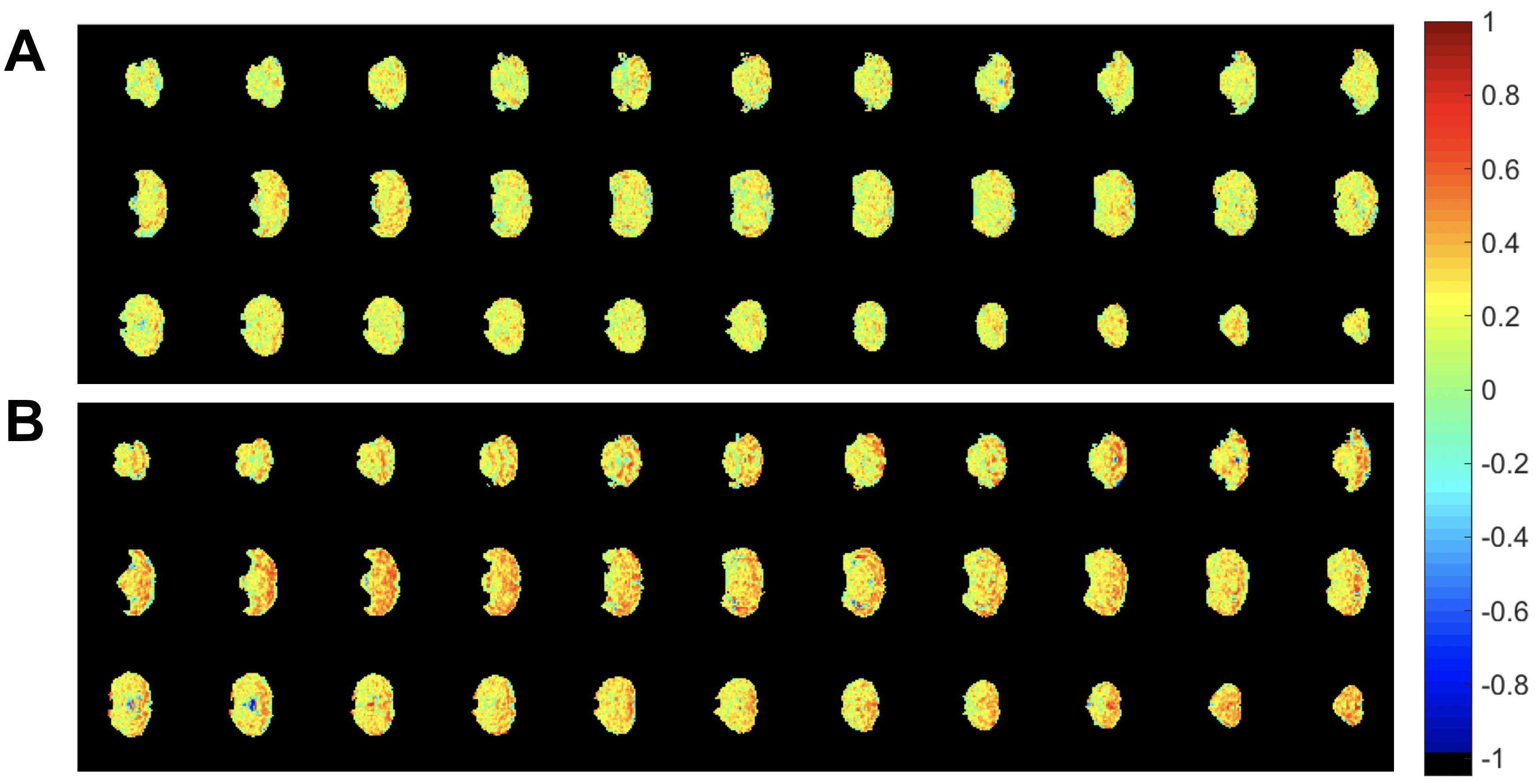
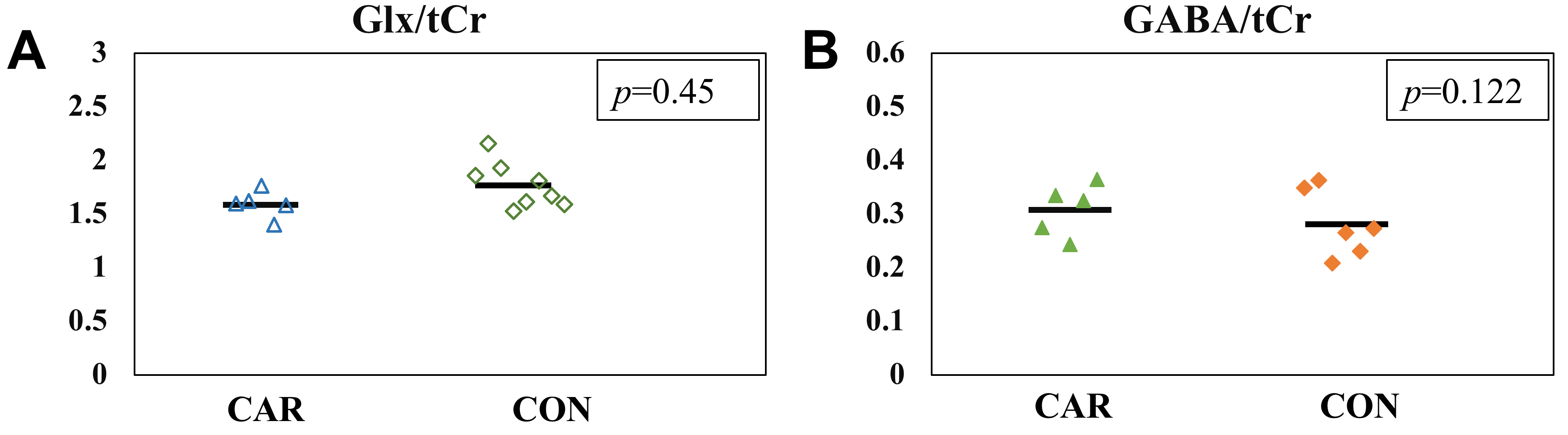
VBM was used to measure the volume of different brain regions, including grey matter (GM), white matter (WM), hippocampus, prefrontal cortex (PFC) and the medial PFC (mPFC). Differences were seen particularly in the mPFC (P = 0.0092, Fig. 1). Compared with CON group, the volume of mPFC is significantly decreased in CAR group. rsfMRI analysis revealed that correlation within the cortex with a seed placed in the mPFC was increased in the CAR group versus the CON group (Fig. 2). This indicated altered functional connectivity in the CAR group. To further investigate the relationship between network-level functional connectivity and neurochemical activity, we measured the concentration of the excitatory neurotransmitter glutamate / glutamine (Glx) and the inhibitory neurotransmitter gamma-aminobutyric acid (GABA) using MRS. Compared with CON group, no significant changes were observed for either Glx or GABA in mPFC in CAR group (Fig. 3). Although studies showed that Glx or GABA is related to resting functional connectivity within brain8,9, the levels of neurotransmitters in mPFC do not represent the whole brain levels. The association of dynamic changes of neurotransmitters in whole brain with the functional connectivity is barely known, which needs further investigation.Conclusion
This work demonstrates that the volume of mPFC is significantly decreased in Rac1 activation group. Moreover, we found that the upregulation of Rac1 activity is related to increased functional connectivity in mouse brain. However, Glx or GABA levels do not show significant changes in mPFC. We concluded that the upregulation of Rac1 activity is related to structural and functional neuroimaging changes.Acknowledgements
This work was supported by grants from the National Natural Science Foundation of China (81873893), the key project of Shanghai Science &Technology (16JC1420402), and Shanghai Municipal Science and Technology Major Project (2018SHZDZX01).References
- Jaffe AB, and Hall A. Rho GTPases: biochemistry and biology. Annu. Rev. Cell Dev. Biol. 2005; 21:247-269.
- Sanz-Moreno V, Gadea G, et al. Rac activation and inactivation control plasticity of tumor cell movement. Cell. 2008; 135 (3): 510–23.
- Heasman SJ., and Ridley AJ. Mammalian Rho GTPases: new insights into their functions from in vivo studies. Nat. Rev. Mol. Cell Biol. 2008; 9:690-701.
- Tejada-Simon MV. J Neurochem. 2015;133(6):767-779.
- Janz P, Savanthrapadian S, Häussler U, et al. Synaptic remodeling of entorhinal input contributes to an aberrant hippocampal network in temporal lobe epilepsy. Cereb Cortex. 2017;27(3):2348-2364.
- Thompson G, Merritt M, Pan WJ, et al. Neural correlates of time-varying functional connectivity in the rat. NeuroImage. 2013; 83:826-836
- Ullmann JFP, Watson C, Janke AL, et al. A segmentation protocol and MRI atlas of the C57BL/6J mouse neocortex. NeuroImage. 2013; 78:196-203.
- Thielen JW, Hong D, Rohani Rankouhi S, et al. The increase in medial prefrontal glutamate/glutamine concentration during memory encoding is associated with better memory performance and stronger functional connectivity in the human medial prefrontal-thalamus-hippocampus network. Hum Brain Mapp. 2018;39(6):2381-2390.
- Stagg CJ, Bachtiar V, Amadi U, et al. Local GABA concentration is related to network-level resting functional connectivity. Elife. 2014;3:e01465.
Figures