3195
Advanced Experimental setup for awake resting-state fMRI in rabbitsNicola Bertolino1, Daniele Procissi1, Craig Weiss2, Quinn C Smith2, and John F Disterhoft2
1Radiology, Northwestern University, Chicago, IL, United States, 2Physiology, Northwestern University, Chicago, IL, United States
Synopsis
Awake imaging in pre-clinical research is challenging due to the MRI sensitivity to motion and animal’s natural reactivity to unfamiliar and loud environment. In this work we present an experimental setup for resting-state fMRI of rabbits and preliminary data collected with it. Our setup relies on the natural tolerance of rabbit to restraint and a home-designed animal fixing cradle equipped with a 3-channel receiver coil.
Introduction
The analysis of functional brain network using resting-state fMRI (rs-fMRI) is extensively employed to study and characterize cognitive impairment present in patients affected by neurodegenerative disease1. Moreover, alteration in functional connectivity seems to occur in early stages of the diseases before the manifestation of gross morphological damage or other obvious neurological synptoms2. For this reason, investigating resting state connectivity in animal models of cognitive impairment offers a precious tool to gain insight into progression of neurodegenerative disease. Due to the sensitivity of MRI to motion artifacts, awake imaging can be challenging in preclinical studies involving animals. Awake imaging is not only necessary for task-based fMRI, but also anesthesia or sedation can alter the BOLD signal and functional connectivity3,4 creating a confounding effect when interpreting group differences. In this perspective, rabbits have been shown to be ideal subjects for awake studies because of their tolerance to restraint5. Using a 3-channel coil specifically designed to enable head-posting and restraint we conducted experiments to advance our previous work6 and to further enhance investigation of resting-state functional networks.Material and Methods
All experimental procedures involving animals complied with Northwestern’s IACUC guidelines. Four Rabbits underwent habituation to MRI environment. Acquisitions were performed on Bruker 7T ClinScan MRI using a three-channel custom made receiver coil (RAPID MR International, Columbus (OH)) designed with a central aperture that allows animal head fixation. Volume coil was used for transmission (Fig1). The animal cradle was designed to integrate the 3-channel receiver coil in a cross-bar fixed to the holder (Fig2). Prior to MRI surgery was performed on each rabbit. Two nylon bolts were fixed on the skull to lock the head into the 3-channel coil limiting head motion during MRI. Each rabbit was inserted in a Lomir Snuggle Sack to reduce stress and then placed in the cradle (Fig3) with the head locked in the cross-bar using the skull fixed bolts. Physiological parameters were recorded throughout the experiment inside the MRI. The acquisition protocol included a coronal 3D-GRE multi-echo scan (TR=68ms; TEs=2.7,6.83,11.26,16,10.13,25ms; Flip-Angle=15; voxel size= 0.29x0.29x0.5mm3; FOV=29.6x55.6x24mm3) and an transverse EPI for rs-fMRI (TR=1800ms; TE=25; Flip-Angle=70; voxel size=0.5x0.5mm2; slice-thickness=1.5; slices=20; FOV= 34x26mm2; GRAPPA=2; echo spacing=0.25ms; volumes=500). The rs-fMRI was repeated twice. Data analysis was performed using FMRIB Software Library v6.0 (Analysis Group, FMRIB, Oxford, UK). A high-resolution 3D image of each brain was generated from the 5 echo average. The 3D images from each rabbit were co-registered (non-rigid 12 degree-of-freedom transformation) and used to generate the rabbit brain template (Fig4) after skull removal. For the resting-state analysis, EPIs were first preprocessed: i)all volumes were registered by a rigid transformation to the central volume, ii)a brain mask was generated by the bias field corrected mean of the volumes and used as an inclusive mask for EPI, iii)a common origin was selected for all subjects’ EPIs, iv)slice timing correction was performed, v)time course was high-pass filtered with a threshold of 0.02 Hz and vi)images were smoothed using a 0.5 mm gaussian kernel. Visual inspection of EPI volumes and motion correction reports enabled overall quality control of data. Functional volumes were registered to high-resolution 3D images and then to the common template before independent components analysis (ICA). ICA group-analysis was run using a multi-session temporal concatenation pre-selecting 40 desired components. Resulting component were finally inspected to identify resting state network and spurious components.Results
The second EPI repetition of one subject was excluded because of excessive body motion. The main functional networks previously described in the literature were identified and are shown in Figure 5.Discussion and Conclusion
Reduction of motion artifacts in awake imaging requires head-posting of animals. This limits the types of rf coils which can be used for imaging. Most experiments employ open single channel surface coils with limited temporal resolution and coverage. To the best of our knowledge this is the first small animal awake rs-fMRI study using a customized phase-array 3 channel coil compatible with head-posting. With this coil we were able to use parallel imaging scheme (GRAPPA acceleration factor) increasing temporal and spatial resolution and extending whole brain coverage. Our setup allowed enhanced localization accuracy and improved temporal characterization of resting state fuctuations7. The ability to the delineate the expected independent components despite the small group of animals verified the good sensitivity of the described setup.Acknowledgements
No acknowledgement found.References
- Van Den Heuvel, Martijn P., and Hilleke E. Hulshoff Pol. "Exploring the brain network: a review on resting-state fMRI functional connectivity." European neuropsychopharmacology 20.8 (2010): 519-534.
- Rombouts, Serge ARB, et al. "Altered resting state networks in mild cognitive impairment and mild Alzheimer's disease: an fMRI study." Human brain mapping 26.4 (2005): 231-239.
- Paasonen, Jaakko, et al. "Functional connectivity under six anesthesia protocols and the awake condition in rat brain." NeuroImage 172 (2018): 9-20.
- Ciobanu, Luisa, et al. "Effects of anesthetic agents on brain blood oxygenation level revealed with ultra-high field MRI." PloS one 7.3 (2012): e32645.
- Weiss, Craig, et al. "The rabbit as a behavioral model system for magnetic resonance imaging." Journal of neuroscience methods 300 (2018): 196-205.
- Schroeder, Matthew P., et al. "Intrinsic connectivity of neural networks in the awake rabbit." Neuroimage 129 (2016): 260-267.
- Zalesky, Andrew, et al. "Time-resolved resting-state brain networks." Proceedings of the National Academy of Sciences(2014): 201400181.
Figures

Fig 1: Image showing the 3-channel receiver
coil placed inside the animal cradle with the two skull-mounted nylon bolts
secured to the cross-bar with nylon nuts.
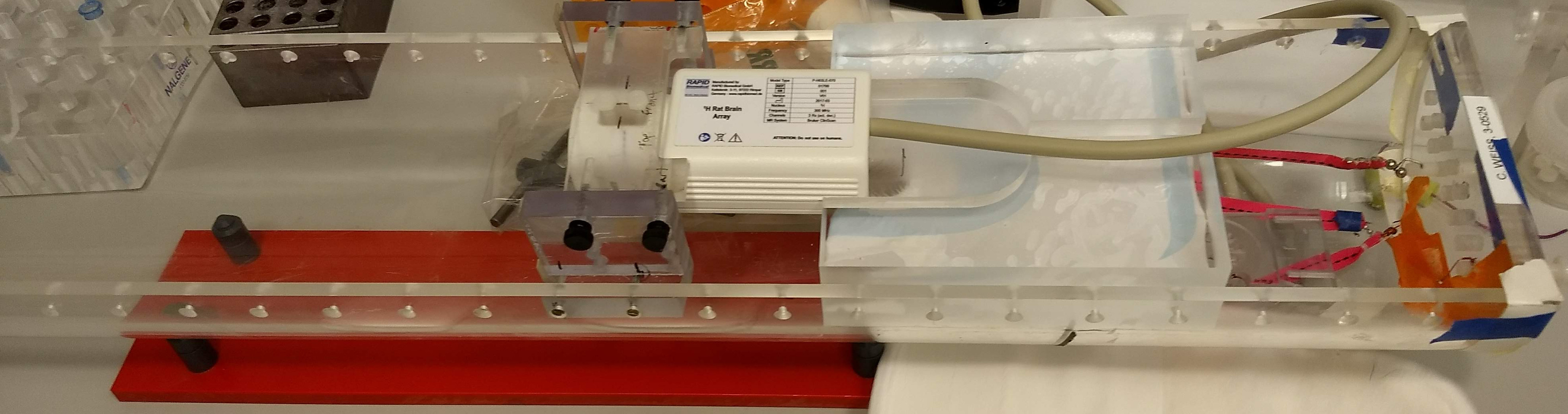
Fig 2: The animal cradle, cross-bar (supporting the far left of the
coil) and an acrylic block to support the body of the pre-amplifier.

Fig 3: A rabbit inside the animal cradle ready
for MRI acquisition. The animal was placed in a cloth sack (blue) and secured
in a canvas Lomir Snuggle Sack.

Fig 4: An image showing representative slices
of the rabbit brain template (dorsal side down).
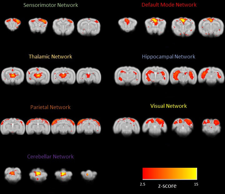
Fig 5: An image showing the result of the ICA
group analysis. The main functional networks described in Schroeder et al.
(2016) were identified.