3188
A novel MRI classifier of arteriolar sclerosis in aging: Prediction of pathology and cognitive decline1Department of Biomedical Engineering, Illinois Institute of Technology, Chicago, IL, United States, 2Rush Alzheimer's Disease Center, Rush University, Chicago, IL, United States, 3Department of Neurological Sciences, Rush University, Chicago, IL, United States, 4Department of Pathology, Rush University, Chicago, IL, United States, 5Department of Diagnostic Radiology, Rush University, Chicago, IL, United States
Synopsis
Arteriolar sclerosis is one of the main pathologies of small vessel disease, is common in the aging brain, and has been associated with lower cognitive performance and higher risk of dementia. Definitive diagnosis of arteriolar sclerosis is only possible at autopsy. In this work, an MRI-based classifier of arteriolar sclerosis was developed, by first training a classifier on ex-vivo MRI and pathology data and then translating it in-vivo, and was evaluated in a large community cohort of older adults.
Introduction
Arteriolar sclerosis is one of the main pathologies of small vessel disease, is common in the aging brain, and has been associated with lower cognitive performance and higher risk of dementia1. Definitive diagnosis of arteriolar sclerosis is only possible at autopsy. In this work, an MRI-based classifier of arteriolar sclerosis was developed, by first training a classifier on ex-vivo MRI and pathology data and then translating it in-vivo. The new classifier was evaluated ex-vivo based on its performance in predicting arteriolar sclerosis, and in-vivo, by testing its association with cognitive decline following baseline in-vivo MRI, in a large community cohort of older adults.Methods
Development of an ex-vivo MRI classifier of arteriolar sclerosis:
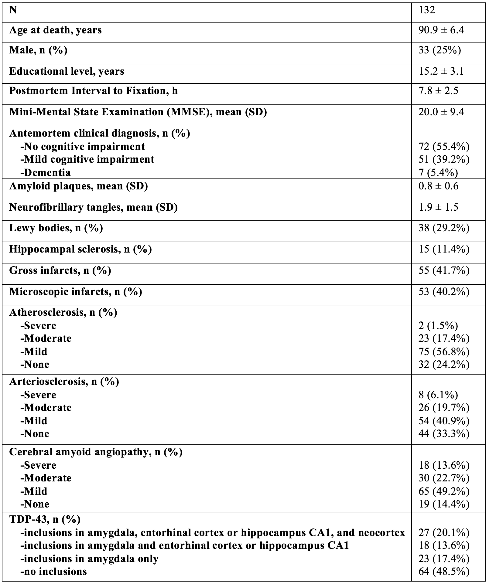
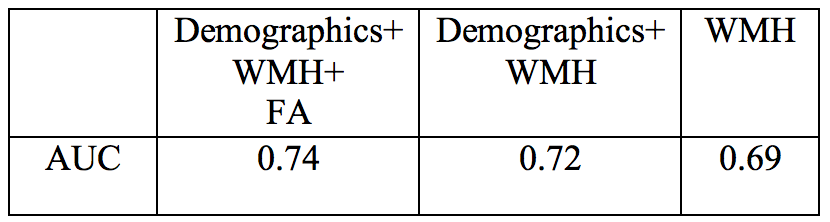
Cerebral hemispheres were obtained from 105 deceased participants of the Rush Memory and Aging Project2 (MAP), and the Religious Orders Study3 (ROS), two longitudinal, clinical-pathologic cohort studies of aging (Fig.1). All hemispheres were imaged ex-vivo on a 3T clinical MRI scanner, while immersed in 4% formaldehyde solution. Following ex-vivo MRI, all hemispheres underwent neuropathologic examination by a board-certified neuropathologist blinded to clinical and imaging findings. Regional white matter hyperintensities (WMH) volumes, and regional fractional anisotropy (FA) values were extracted from the ex-vivo MRI data and used as features in a linear support vector machine classifier of arteriolar sclerosis. Other features included basic demographic information (i.e. age, sex, years of education). Performance evaluation involved 100 repeats of stratified shuffle split cross-validation with 80% of the data used for training, and 20% for testing. The process was repeated for a classifier based exclusively on WMH, which historically have been considered a marker of small vessel disease4,5. The performance of the two classifiers was compared using a Wilcoxon signed rank test (p<0.05).
In-vivo translation of the classifier:
Both in-vivo and ex-vivo 3T MRI data were collected on 13 participants. Regional WMH and FA values were extracted from the in-vivo and ex-vivo MRI data and linear mixed-effects models were used to establish the relationship between the in-vivo and ex-vivo values. These models were used to achieve in-vivo translation of the classifier.
Testing the classifier in-vivo:
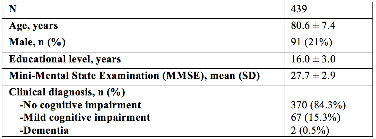
In-vivo 3T MRI and clinical data were obtained on 439 MAP and ROS participants (Fig.2). For each participant, regional WMH and FA values were extracted from the in-vivo MRI data, were converted to ex-vivo values using the models from the previous section, were entered into the classifier, and a classification confidence score was generated. This classification confidence score expressed the likelihood that a participant suffered by arteriolar sclerosis. Since pathology information was not available for this large cohort of older adults, the performance of the classifier in-vivo was assessed by testing the association of the classification confidence score with change in global cognition two years after baseline MRI, using Pearson’s correlation. The same analysis was repeated for 5 cognitive domains: semantic memory, episodic memory, working memory, perceptual speed, and visuospatial abilities.
Results
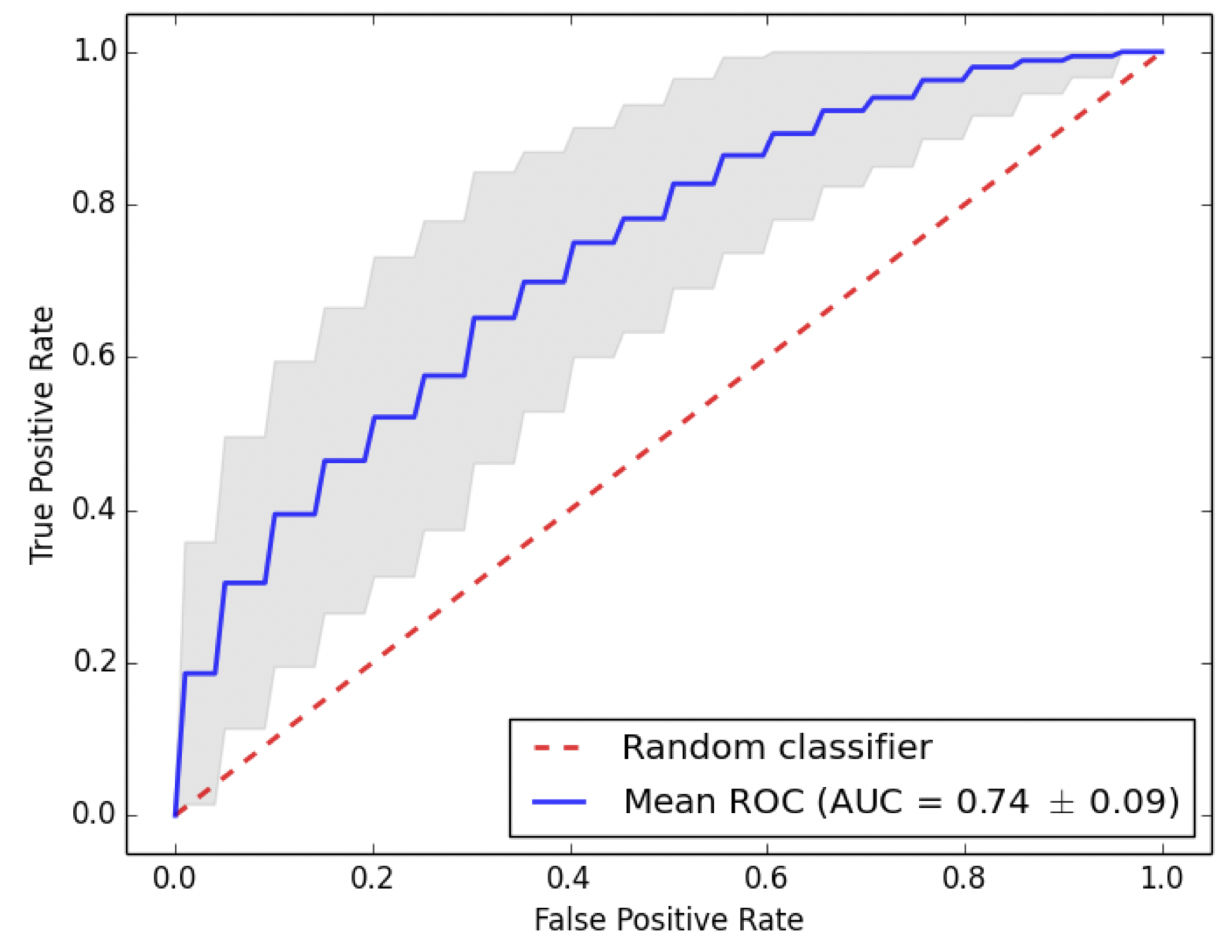
The average area under the receiver operating characteristic curve (AUC) for ex-vivo classification of arteriolar sclerosis was 0.74 for the classifier based on WMH, FA, and demographics (Fig.3), which was higher than that of the classifier based exclusively on WMH (0.69) (p<0.05) (Fig. 4). The cohort used for testing the classifier in-vivo was on average 10 years younger than the cohort used for training the classifier (ex-vivo) (Figs.1,2). After translating the classifier in-vivo, the classification confidence score was associated with two-year decline in perceptual speed (beta=-0.17, p=0.0004) and visuospatial abilities (beta=-0.11, p=0.02), and two-year decline in global cognition (beta=-0.14, p=0.003).Discussion
A novel MRI-based classifier of arteriolar sclerosis was developed based on ex-vivo MRI and pathology data and tested both ex-vivo and in-vivo. Ex-vivo testing demonstrated that the new classifier has superior performance in predicting arteriolar sclerosis compared to a classifier based exclusively on WMH. In-vivo translation and testing in a large community cohort, showed that the new classifier is associated with cognitive decline in two years after baseline MRI. This finding illustrates the clinical relevance of the new classifier. Furthermore, the fact that the cohort used for in-vivo testing was on average 10 years younger than the cohort used for ex-vivo training of the classifier, shows that the classifier generalizes to in-vivo data collected earlier in life. The fact that the classification confidence score was most strongly correlated with two year decline in perceptual speed is particularly important as small vessel disease is known to have a strong effect on this cognitive domain.Conclusion
A novel MRI-based classifier was developed for arteriolar sclerosis, a neuropathology that can only be diagnosed at autopsy.Acknowledgements
UH2-UH3 NS100599References
1. Arvanitakis Z, Capuano AW, Leurgans SE, Bennett DA, Schneider JA. Relation of Cerebral Vessel Disease to Alzheimer’s Disease Dementia and Cognitive Function in Older Persons: A Cross-sectional Study. The Lancet Neurology. 2016;15(9):934-943.
2. Bennett DA, Schneider JA, Buchman AS, et al. Overview and findings from the Rush Memory and Aging Project. Curr Alzheimer Res 2012;9:646–663.
3. Bennett DA, Wilson RS, Arvanitakis Z, et al. Selected findings from the Religious Orders Study and Rush Memory and Aging Project. J Alzheimers Dis 2013;33 Suppl 1:S397-403.
4. Debette S, Markus HS. The clinical importance of white matter hyperintensities on brain magnetic resonance imaging: systematic review and meta- analysis. BMJ 2010;341:c3666.
5. Erten-Lyons D, Dodge HH, Woltjer R, et al. Neuropathologic basis of age-associated brain atrophy. Neurology 2013;70:616-622.
Figures