3179
Functional Impact of Theta Burst Stimulation on Motor Cortex1Biomedical Engineering, University of Arizona, Tucson, AZ, United States, 2Psychology, University of Arizona, Tucson, AZ, United States
Synopsis
The effect of transcranial magnetic stimulation using intermittent theta burst stimulation (iTBS) on functional connectivity and cortical excitability was tested in a healthy older adult and individuals with mild cognitive impairment. Individuals underwent an iTBS protocol with single pulse cortical excitability tests and-resting state fMRI(rs-fMRI) being performed before and after. Cortical excitability was tested through a pseudorandomized order of intensities of single pulses. Within functional network RS-fMRI analysis was done of the sensorimotor network. Differences in the response between the two populations was observed in both the functional connectivity changes in the sensorimotor network as well as the cortical excitability.
Introduction
Transcranial magnetic stimulation (TMS) is a noninvasive method to stimulate the brain through the use of rapidly alternating magnetic fields that can induce electrical currents in the brain. Depending on the frequency of TMS pulses applied, the effect on the stimulated brain tissue can be either excitatory or inhibitory. Theta Burst Stimulation (TBS) is a form of repetitive TMS that is considered the most efficient protocol in terms of number of impulses and intensity required during a given stimulation1. In this study, we aimed to study the effect of intermittent TBS (iTBS, excitatory stimulation) on functional connectivity and cortical excitability in healthy older adults as well as individuals with mild cognitive impairment (MCI).Methods
14 healthy older adults (7 male, age 69.4±2.9) who were age matched to 4 individuals with MCI were enrolled in this study. The TMS protocol comprised of interleaved single-pulse TMS and iTBS over the left primary motor cortex. Measures of cortical excitability were acquired from the contralateral abductor pollicis brevis (APB) with surface EMG immediately before and after the iTBS protocol. The primary cortical excitability outcome measures were obtained with a stimulus response curve, which was acquired in an active condition in the presence of a voluntary tonic muscle contraction of the APB. The single-pulse TMS protocol comprised of 64 pulses, which were delivered in a pseudorandomized order and ranged in intensity from 80-150% of the individuals active motor threshold (AMT). The standard iTBS protocol comprised of 600 pulses1.
The imaging protocol was comprised of two imaging sessions, immediately proceeding and following the iTBS. Imaging was performed on a 3T scanner (Siemens, Skyra). Structural T1 weighted images using an MPRAGE sequence were acquired for registration with TMS Navigation System. Echo-planar resting state functional MRI images were acquired with TR/TE=3000/36 msec, FOV=240mmx240mmx108mm, FA= 90, acquisition matrix=160x80x72, voxel size=1.5mm3, and multi-band factor=2. Data was preprocessed using Matlab, SPM and CONN Toolbox. Motion and slice timing correction were performed using SPM.
Independent component analysis was done on the data set to
ensure that the image quality was consistent.
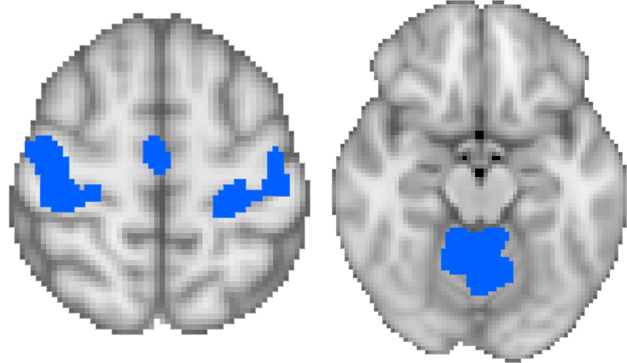
Further analysis was performed through correlation matrix analysis to
look at within functional network correlations for the stimulated network
(i.e., the sensorimotor network) (Figure 1). This
was done by determining a Fisher r-to-z transformed Pearson correlation
coefficient between each ROI within the functional network of interest2,
then taking an average of the correlation values for each network. Paired t-tests were performed to determine if
there were statistically significant changes in network connectivity before and
after the iTBS protocol. Furthermore, results
were compared between the two population groups to determine if there were differences
in response to iTBS.
Results
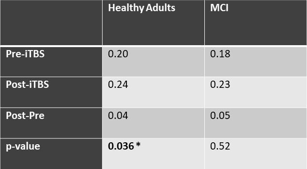
Analysis of within-network correlation showed that functional connectivity within the sensorimotor network significantly increased after the iTBS in healthy older adults (p = 0.036) (Table 1). Motor-evoked potential as measured by the EMG also significantly increased after the iTBS in healthy older adults, F(1,12) = 4.91, p = 0.047, indicating an excitatory effect. Due to the small sample size in the MCI group, currently, no significant changes in the sensorimotor network were observed (p > 0.05).Discussion
The results indicate that iTBS increased both motor-evoked potential as well as resting-state functional connectivity within the sensorimotor network in healthy older adults. Our study suggests that the enhanced cortical excitability induced by iTBS parallels a more coherent sensorimotor network. This finding is in line with previous animal studies indicating long-term potentiation like effects underlying iTBS aftereffects. The recruitment for the MCI population is ongoing. Further studies of clinical populations investigating mechanisms underlying iTBS-induced changes in cortical excitability and plasticity will be needed.Acknowledgements
Support by NIH P30 AG019610 Arizona Alzheimer’s Consortium Pilot Study Program and University of Arizona BIO5 Team Scholars Award.References
1. Huang Y-Z, Edwards MJ, Rounis E, Bhatia KP, Rothwell JC. Theta Burst Stimulation of the Human Motor Cortex. Neuron [Internet] 2005;45:201–206. doi: 10.1016/j.neuron.2004.12.033.
2. Shirer WR, Ryali S, Rykhlevskaia E, Menon V, Greicius MD. Decoding Subject-Driven Cognitive States with Whole-Brain Connectivity Patterns. Cereb. Cortex [Internet] 2012;22:158–165. doi: 10.1093/cercor/bhr099.
Figures