3178
HIGH AND LOW RISK DEMENTIA CLASSIFICATION IN EARLY MCI FROM ADNI11Cognitive Neuroscience, Center for Mind/Brain Sciences, University of Trento, Rovereto, Italy, 2Neuroradiology, Santa Maria del Carmine Hospital of Rovereto, Rovereto, Italy, 3Center for Mind/Brain Sciences, University of Trento, Mattarello, Italy
Synopsis
Mild cognitive impairment (MCI) has been defined as the stage between the expected cognitive decline of normal aging and the more serious decline of dementia. Its clinical characteristics represent the earliest features of many forms of dementia. Specifically, many studies have reported amnestic MCI (aMCI) as a risk factor for Alzheimer’s disease, but to date there’s still a need for establishing specific, reliable, pre-symptomatic and non-invasive biomarkers associated to the progression of aMCI to AD. In this study we investigated CSF and structural MRI biomarkers for the classification of high and low risk of dementia conversion in aMCI.
INTRODUCTION
A recent European multicentric study performed by Marizzoni et. al1, found that in amnestic MCI with similar early cognitive deficits, the ratio of CSF-derived amyloid beta and p-tau can help stratify low- and high-risk groups of conversion. The same study found that, from a series of structural (MRI morphometry and diffusion) and functional (resting state fMRI, resting state and odd-ball EEG) data9, the most sensitive marker to AD progression was the segmented volume of the lateral ventricles and medial temporal lobe (p≤0.042). This study, however, was based on the use of 3T MRI. Thus, with an interest of testing the validity and application value of their results on aMCI patients scanned at 1.5T in the Hospital of Rovereto (Italy), we first investigated these biomarkers in aMCI patients from the public longitudinal ADNI dataset obtained with a 1.5T MRI system. Particularly, the CSF derived amyloid beta and p-tau, ventricular volume segmentations and the ADASCOG 13 scores were considered for the evaluation of low- and high-risk groups as reported by Marizzoni et al.MATERIALS & METHODS
112 ADNI1 patients were selected based on their screening time (baseline, 6months and 12 months), CSF values (i.e. amyloid-β (Aβ) and p-tau), FSL calculated left and right ventricular and hippocampal volumes (using 1.5T MRI systems), and ADAS-Cog 13 scores across all three time points. For the stratification of risk levels, a two-way gaussian mixed model was created using MATLAB software version R2018a to model the baseline values of amyloid-β/p-tau ratios and baseline lateral ventricular volumes of the selected patients. Lastly, a mixed analysis of variance (ANOVA) was performed using SPSS statistics on the subpopulations produced by each model for the longitudinal assessment of cognition and atrophy. Age and gender were chosen as nuisance variables.RESULTS
The baseline amyloid-β/p-tau gaussian mixed model produced a cut off value of 34 pg/mL with 72 high-risk patients (below cut off) and 40 low-risk patients (above cut off), with means and standard deviations of: µ1 = 63.5, σ1 = 467.6, µ2 = 18.8, σ2 =47.7 respectively. In contrast, the baseline ventricular volume mixed model produced a cut off volume of volume of 70.5 mm3 with 102 low-risk patients (below cut off) and 10 high-risk patients (above cut off). With means and standard deviations of: µ1 = 89240 σ1 = 8 e+08, µ2 = 36125, σ2 = 2 e+08. ANOVA results reveal that patients classified as high-risk by both biomarkers had worse cognitive decline over time (i.e. higher ADAS-Cog13 scores) and smaller hippocampal volumes. In contrast, patients classified as low-risk by both markers had better cognitive function (i.e. lower ADAS-Cog13 scores) and larger hippocampal volumes over time.DISCUSSION
Our study reveals that aMCI patient with lower amyloid-β/p-tau ratios and larger ventricular volumes are at a higher risk of cognitive decline that could consequently lead to AD dementia. These results are consistent with those reported by Marizzoni et. al1 and other longitudinal studies 2-7 that have tested the same hypothesis on large and small sample sizes of AD, MCI and healthy controls. The Aβ42/P-tau ratio derived from CSF has been reported as a promising parameter for discriminating between MCI patients with high risk of conversion from MCI stable patients with a sensitivity of 81% and specificity of 95%8. However, due to the invasiveness of the lumbar puncture procedure, the use of CSF biomarkers is significantly limited in daily clinical practice. Thus, the use of structural biomarkers such as the lateral ventricular volumes together with other structural biomarkers (e.g. hippocampus, amygdala, periventricular volumes) could be a less invasive alternative for the early prediction of disease progression in aMCI.CONCLUSION
This study extends previous 3T MRI study to the ADNI1 data (1.5T MRI) providing additional evidence that the lateral ventricular volume and amyloid-β/p-tau ratios are good candidate biomarkers for the prediction of disease progression in aMCI patients. However, in cases where CSF biomarkers are not available structural MRI markers could be investigated collectively to increase specificity and sensitivity for prediction of disease progression. Lastly, we envision that the next step in clinical research could be the creation of standardized procedures for the proper use of these markers.Acknowledgements
No acknowledgement found.References
1. Marizzoni, Moiraa, Ferrari, Clarissa, Jovicich, Jorgec, Albani, Diegod, Babiloni, Claudioe; Cavaliere, Liberaa, Didic, Mirag; h, Forloni, Gianluigid, Galluzzi, Samanthaa, Hoffmann, Karl-Titusi, Molinuevo, José Luisj, Nobili, Flaviok et al. (2018). Predicting and Tracking Short Term Disease Progression in Amnestic Mild Cognitive Impairment Patients with Prodromal Alzheimer’s Disease: Structural Brain Biomarkers. Journal of Alzheimer’s Disease.
2. C. R. Jack, M. M. Shiung, J. L. Gunter, P. C. O’Brien, S. D. Weigand, D. S. Knopman, B. F. Boeve, R. J. Ivnik, G. E. Smith, R. H. Cha, E. G. Tangalos, R. C. Petersen. (2004). Comparison of different MRI brain atrophy rate measures with clinical disease progression in AD. Neurology .
3. David AvilaEmail the author David Avila, Sona Hurtz, Anna Blanken, Kristy Hwang, Giovanni Coppola, Liana Apostolova. (2014). Effects On Ventricular Enlargement When Comparing Cognitively Normal Elderly, Mild Cognitive Impairment With Alzheimer's Disease Subjects. . The Journal of the Azlheimer's Association. .
4. JieShi, Cynthia M.Stonnington, Paul M.Thompson, KeweiChendBorisGutman, ColeReschke, Leslie C, BaxtereEric M.Reiman, Richard J.Caselli, YalinWang, for the Alzheimer's Disease Neuroimaging Initiative. (2014). Studying ventricular abnormalities in mild cognitive impairment with hyperbolic Ricci flow and tensor-based morphometry. NeuroImage Volume 104, 1 January 2015, Pages 1-20.
5. Kelvin K. Leung, Jonathan W. Bartlett, Josephine Barnes, Emily N. Manning, Sebastien Ourselin, Nick C. Fox, for the Alzheimer's Disease Neuroimaging Initiative. (2013). Cerebral atrophy in mild cognitive impairment and Alzheimer disease. Neurology .
6. W.J.P. Henneman, H. Vrenken, J. Barnes, I. C. Sluimer, N. A. Verwey, M. A. Blankenstein, M. Klein, N. C. Fox, P. Scheltens, F. Barkhof, W. M. van der Flier. (2009). Baseline CSF p-tau levels independently predict progression of hippocampal atrophy in Alzheimer disease. Neurology.
7. Yi-Yu Chou, Natasha Leporé, Priya Saharan, Sarah K. Madsen, Xue Hua , Clifford R. Jack et. al. (2010). Ventricular maps in 804 subjects correlate with cognitive decline, CSF pathology, and imminent Alzheimer's disease. IEEE International Symposium on Biomedical Imaging: From Nano to Macro.
8. Parnetti, Lucillaa, Chiasserini, Davidea,Eusebi, Paolo, Giannandrea, Davida, Bellomo, Giannic, De Carlo, Claudiaa, Padiglioni, Chiaraa et. al. (2012). Performance of Aβ1-40, Aβ1-42, Total Tau, and Phosphorylated Tau as Predictors of Dementia in a Cohort of Patients with Mild Cognitive Impairment. : Journal of Alzheimer's Disease, vol. 29, no. 1, pp. 229-238, 2012.
9. Jovicich Jorge, Babiloni, Claudio, Ferrari, Clarissa, Marizzoni, Moira, Moretti, Davide V, Del Percio, Claudiog, Lizio, Robertab et. al. (October 2018 ). Two-Year Longitudinal Monitoring of Amnestic Mild Cognitive Impairment Patients with Prodromal Alzheimer’s Disease Using Topographical Biomarkers Derived from Functional Magnetic Resonance Imaging and Electroencephalographic Activity. Journal of Alzheimer's Disease , vol. Pre-press, no. Pre-press, pp. 1-21, 2018.
Figures
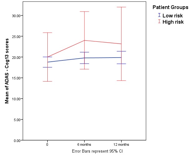
Figure 1. Illustrates the mean ADAS-Cog scores of each patient group across three time points. Patients with smaller ventricular volumes were classified as low risk of disease progression and patients with larger ventricular volumes were classified high risk. Statistical analysis reveals no interaction effect (Wilk’s lambada = .980, F = (2,109) = 1.126, p = .328, η 2 = .020) and no significant effect of time (Wilk’s lambada = .946 , F = (2,109) = 3.114 , p = .048 η 2 = .054), suggesting that the difference in ADAS-Cog measurements was mainly due to the difference in patient groups.
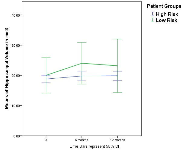
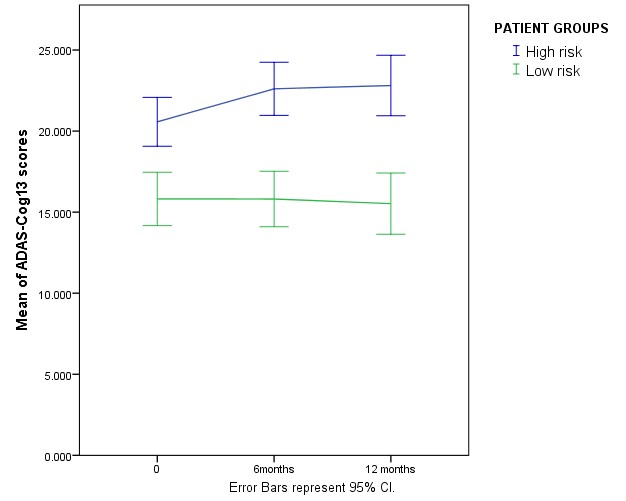
Figure 3. Illustrates the mean ADAS-Cog scores of patient groups as classified by the amyloid-β/p-tau ratios. Statistical analysis reveals no interaction effect (Wilk’s lambada = .944, F = (2,111) = 3.316, p = .040, η 2 = .056) and no significant effect of time (Wilk’s lambada = .957, F = (2,111) = 2.523, p = .085 η 2 = .043).These suggests that the difference in ADAS-Cog measurements was mainly due to the difference in patient groups (p = .000).