3166
White matter hyperintensity burden is strongly associated with neuro-inflammation but not with amyloid deposition: a multimodal PET MR studyHongyu An1, Chunwei Ying1, Yasheng Chen2, Peter Kang2, Jon Christensen1, Qing Wang1, Lisa Cash1, Jin-Moo Lee2, Andria Ford2, and Tammie Benzinger1
1Radiology, Washington University in St. Louis, St. Louis, MO, United States, 2Neurology, Washington University in St. Louis, St. Louis, MO, United States
Synopsis
Selectively elevated PET 11C-PK11195 uptake within white matter demonstrates that
Introduction
White matter hyperintensities (WMH) in Fluid Attenuated Inversion Recovery (FLAIR) images have been widely observed in patients with cerebral small vessel disease (CSVD), Alzheimer’s disease (AD), or a mixed CSVD and AD pathology. WMH is strongly associated with progression to dementia. Thus far, the pathogenesis of WMH has been poorly understood. Post-mortem histological studies showing activated microglia, and the presence of inflammatory markers in the blood (CRP) and CSF (matrix metalloproteases and albumin).59,60 More recently, increased and persistent systemic inflammation (measured by CRP) in midlife was found to promote late-life white matter structural injury.36,37 However, there is no in vivo data from patients to provide direct evidence. In addition, because of the high prevalence of mixed CSVD and AD pathology in elderly with WMH, it is unclear whether amyloid deposition, a hallmark early pathological features of AD, may also contribute to WMH. In this study, we sought to evaluate whether WMH burden is associated with neuro-inflammation and amyloid deposition using PET and MR imaging. Moreover, we examined whether neuro-inflammation elevation is region specific.Material & Methods
11C-PK11195 radiotracer binds to activated microglia. Thus it can be used as a molecular imaging biomarkers to measure in vivo inflammation using PET. 19 elderly subjects (12 females, age: 75 [68, 82] (Median [interquartile range IQR])) with various degrees of WMH were recruited and underwent serial PET and MR scans. 11C-PK11195 PET images were acquired to estimate neuro-inflammation. In addition, 11C-PiB PET images, to measure amyloid deposition, were acquired 9-24 months (19 [14, 22] (Median [IQR]) prior to the PET 11C-PK11195 scans from the same patients. WMH lesions were manually outlined using MR FLAIR images and WMH volume is then computed. Moreover, MR T1w MPRage images were used to segment brain tissue into gray matter, white matter and CSF. To account for inter-subject variations in brain tissue volumes, relative WMH (rVWMH) is calculated as a ratio between absolute WMH volume and a summation of gray matter and white matter volumes. All PET and MR images were registered to a FreeSurfer atlas. Median standardized uptake value (SUV) of the cerebellum gray matter was utilized to compute SUV Ratio (SUVR) maps.Results
5 out of 18 patients had a clinical dementia rating (CDR) score greater than 0.5 (CDR=0.5, n=3, CDR=1, n=2). The median and IQR WMH lesion volumes were 8.4 ml [3.6ml 24.1ml]. We found that white matter 11C-PK11195 uptake was significantly higher in subjects with high WMH burden compared to subjects with low WMH burden N=8), while GM uptake did not differ (Fig 1). Univariate linear regression demonstrates that WMH burden is significantly associated with 11C-PK11195 uptake in white matter (Fig 2A, R=0.55, P<0.01) as a weaker association with age (Fig 2B, R=0.51, P<0.05). After adjusting for age, PK uptake shows a near-significant association with WMH burden (Fig 2D, R=0.6, P=0.08). Interestingly, there was no correlation between WMH volumes and amyloid deposition as measured by 11C-PiB PET (Fig 2C, R=-0.17, P=0.50), or with CDR score (data not shown, R = 0.3061 and P = 0.11).Discussion
Translocator protein (TSPO) is expressed on microglia and expression is strongly upregulated upon microglial activation. Autoradiography combined with immunohistochemical staining demonstrate that PK11195 binds to activated microglia in human postmortem brains and in animal models of neurological diseases, thus providing an imaging tool to measure in vivo inflammation.62,63 PET 11C-PK11195 tracer binds to activated microglia and provides a means for imaging neuroinflammation in living humans.60 We have demonstrated that increasing white matter 11C-PK11195 uptake correlate with an increase in WMH volume suggesting the presence of increased inflammation as an index of disease severity. In contrast, PiB uptake, a biomarker of amyloid deposition, is not associated with WMH.Conclusions
Our results demonstrated that neuro-inflammation is selectively elevated in the white matter and significantly correlates with WMH burden. On the other hand, amyloid deposition is not associated with WMH.Acknowledgements
No acknowledgement found.References
1. Rosenberg GA. Extracellular matrix inflammation in vascular cognitive impairment and dementia. Clin Sci (Lond) 2017; 131(6): 425-37. 2. Evans NR, Tarkin JM, Buscombe JR, Markus HS, Rudd JHF, Warburton EA. PET imaging of the neurovascular interface in cerebrovascular disease. Nat Rev Neurol 2017; 13(11): 676-88. 3. Walker KA, Power MC, Hoogeveen RC, et al. Midlife Systemic Inflammation, Late-Life White Matter Integrity, and Cerebral Small Vessel Disease: The Atherosclerosis Risk in Communities Study. Stroke 2017; 48(12): 3196-202. 4. Walker KA, Windham BG, Power MC, et al. The association of mid-to late-life systemic inflammation with white matter structure in older adults: The Atherosclerosis Risk in Communities Study. Neurobiol Aging 2018; 68: 26-33. 5. Banati RB, Newcombe J, Gunn RN, et al. The peripheral benzodiazepine binding site in the brain in multiple sclerosis: quantitative in vivo imaging of microglia as a measure of disease activity. Brain 2000; 123 ( Pt 11): 2321-37. 6. Vowinckel E, Reutens D, Becher B, et al. PK11195 binding to the peripheral benzodiazepine receptor as a marker of microglia activation in multiple sclerosis and experimental autoimmune encephalomyelitis. J Neurosci Res 1997; 50(2): 345-53.Figures
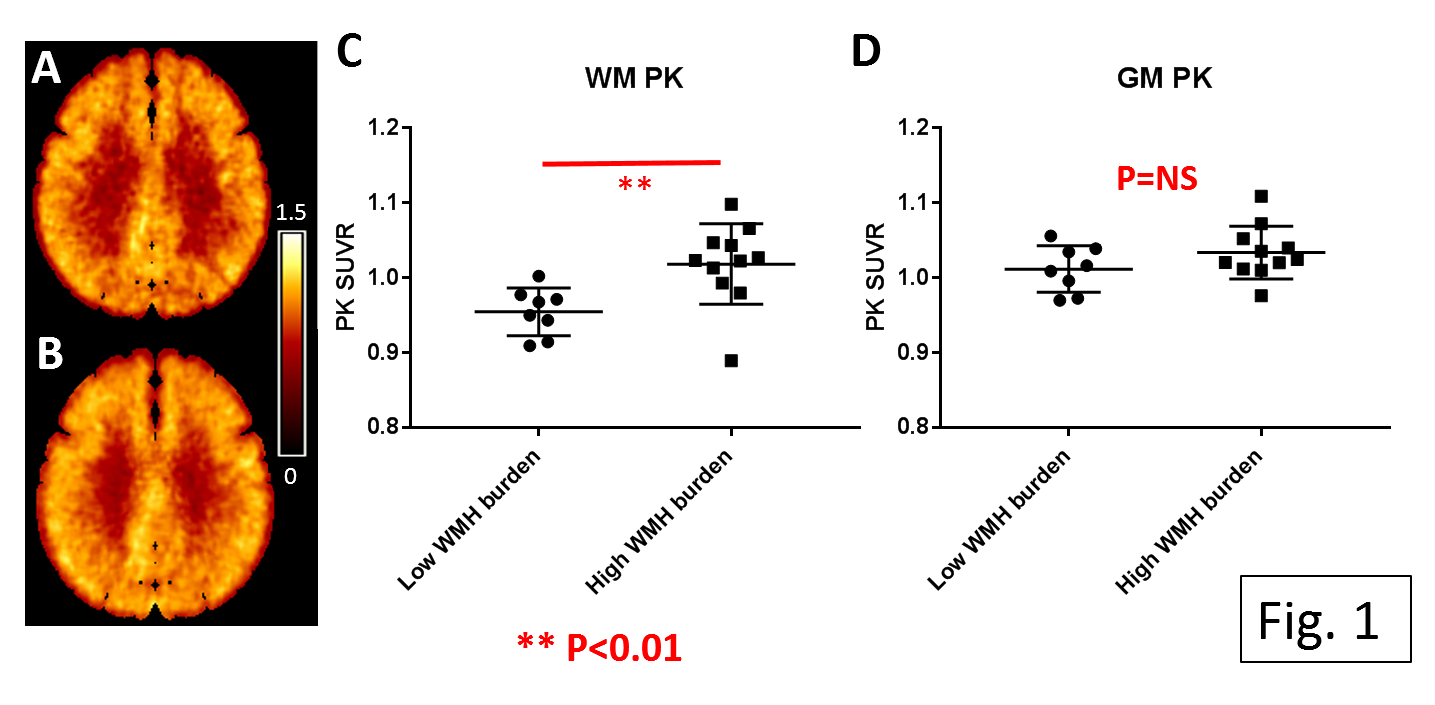
Fig 1. Average PK in low (A , rVWMH≤ 1%) ) and high (B, rVWMH> 1%) WMH burden groups. PK uptake are significantly higher in the high WMH burden group within WM (C), but not within GM (D). The PK uptake difference between the low and high WMH burden groups are 6.7% and 2.2% in WM (P=0.0083) and GM (P=0.172), respectively. ** P<0.01.
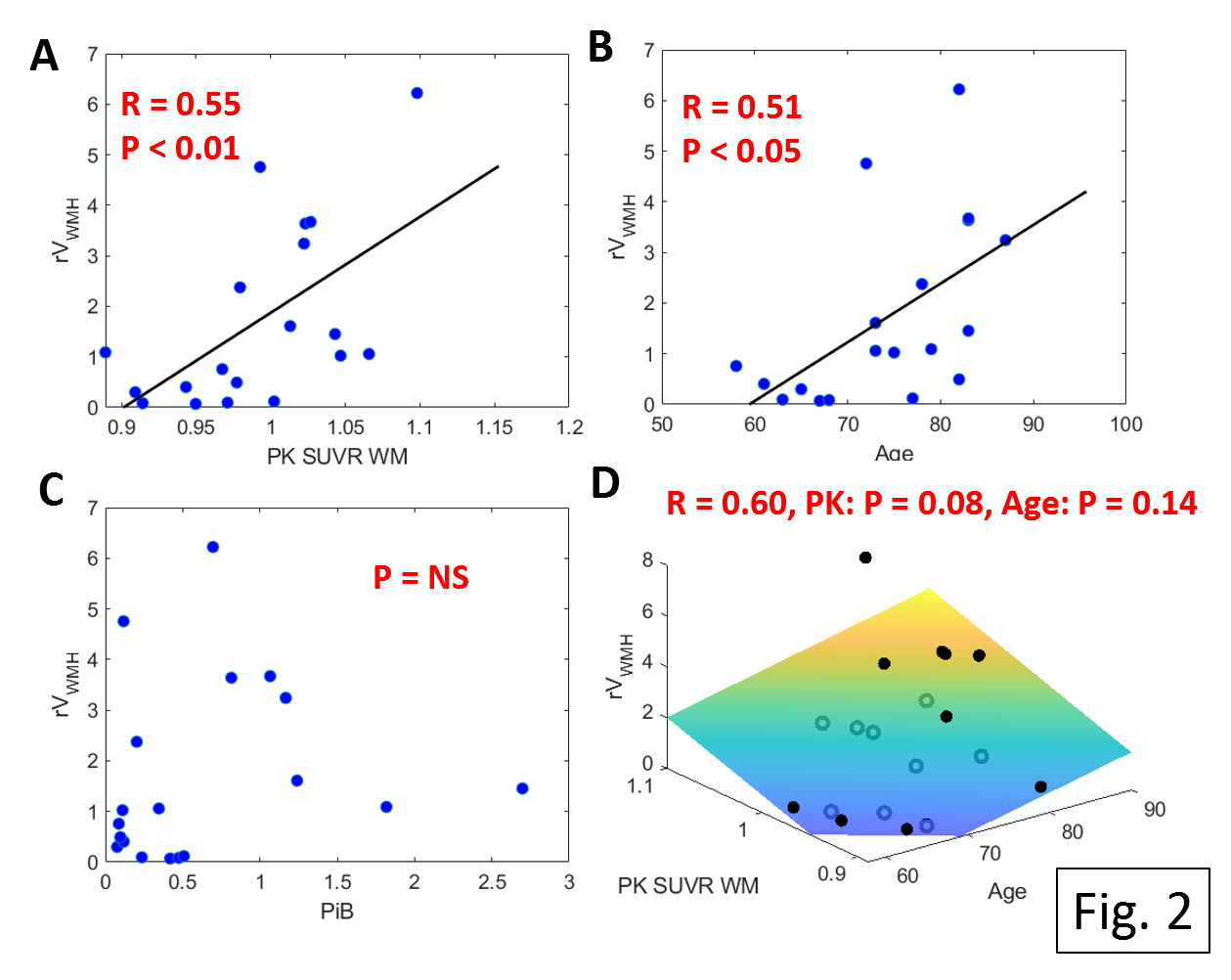
Fig 2. Both PK uptake (A) and age (B) are significantly associated with rVWMH, however there is no correlation between PiB binding potential (C) and rVWMH. PK uptake is near significantly associated with rWMH volume after adjusting for age (D).