3141
In-vivo mapping of thalamic pathological mechanisms in pediatric patients with MSLoredana Storelli1, Ermelinda De Meo1,2, Lucia Moiola2, Maria Pia Amato3, Angelo Ghezzi4, Pierangelo Veggiotti5,6, Ruggero Capra7, Maria A. Rocca1,2, and Massimo Filippi1,2
1Neuroimaging Research Unit, Institute of Experimental Neurology, Division of Neuroscience, San Raffaele Scientific Institute, Vita-Salute San Raffaele University, Milan, Italy, 2Department of Neurology, Institute of Experimental Neurology, Division of Neuroscience, San Raffaele Scientific Institute, Vita-Salute San Raffaele University, Milan, Italy, 3Department NEUROFARBA, Section Neurosciences, University of Florence, Florence, Italy, 4Multiple Sclerosis Center, Ospedale di Gallarate, Gallarate, Italy, 5Department of Child Neurology and Psychiatry, C. Mondino National Neurological Institute, Pavia, Italy, 6Brain and Behaviour Department, University of Pavia, Pavia, Italy, 7Multiple Sclerosis Center, Spedali Civili of Brescia, Brescia, Italy
Synopsis
Despite widely recognized in pediatric population with multiple sclerosis (MS), the pathogenesis of thalamic damage remains largely unknown. This study was performed to explore the microstructural abnormalities within this structure through the use of quantitative MRI metrics (diffusion tensor, T1/T2-weighted ratio) considering the two different thalamic interfaces (cerebrospinal fluid/thalamus and thalamus/white matter) as sites of two different pathogenic processes: the first one accounting for cerebrospinal fluid-mediated factor damage and the second one for diffuse neurodegenerative damage. The study demonstrated an heterogeneous pathogenesis for thalamic damage since the beginning of the disease.
Introduction
Both local inflammatory demyelination and changes secondary to axonal transection of fibers passing through focal white matter (WM) lesions can account for thalamic abnormalities in multiple sclerosis (MS) patients.1 The thalamus plays a major role in brain functioning, integrating inputs originating from the cortex and subcortical structures. As a consequence, thalamic damage has been related to clinical disability and cognitive impairment.2 Assessing location of microstructural abnormalities within the thalamus as a function of distance from cerebrospinal fluid (CSF) in pediatric MS patients could help to identify whether at the beginning of disease, thalamic damage is due to CSF-mediated factors or thalamic neurodegeneration associated with macroscopic damage. The aim of this study is to investigate, in-vivo, pathological mechanisms underlying microstructural thalamic damage in pediatric MS patients by applying quantitative MRI techniques.Methods
Sixty-eight pediatric MS patients and 24 age and sex-matched healthy controls (HC) underwent 3T MRI and clinical evaluation. As quantitative MRI metrics, we assessed diffusion tensor (DT) imaging measures such as fractional anisotropy (FA) and mean diffusivity (MD). In addition, T1/T2-weighted ratio maps were estimated using an in-house implemented method (Matlab®) adapted from Ganzetti et al.3 We tested for: differences in thalamic volume (estimated with FIRST tool from FSL library), and quantitative DT and T1/T2-weighted ratio MRI measures both globally and within concentric bands originating from CSF/thalamus interface (as a function of the geodesic distance of the thalamus from the CSF, Figure 1). Moreover, we assessed the relation between thalamic, cortical, and WM metrics, and the contribution of MRI metrics to clinical disability.Results
Compared to HC, pediatric MS patients had reduced thalamic FA (p=0.009) and no thalamic atrophy. In pediatric MS, significant reduction of FA was observed in bands nearest to CSF and in those nearest to WM compared to HC (Figure 2). Significant abnormalities of MD and T1/T2-weighted ratio were respectively observed in thalamic regions next to CSF and to WM (Figure 3). Global and regional FA abnormalities as well as T1/T2-weighted ratio abnormalities showed significant correlations with disease duration, while MD alterations showed significant correlations with cortical thinning. No significant correlations between thalamic damage and T2 lesion volumes or clinical disability were observed.Discussion
This study demonstrated a heterogeneous pathogenesis for thalamic damage since the beginning of the disease, accounting for both cerebrospinal fluid mediated and diffuse normal appearing WM damage.Conclusions
The abnormalities observed suggest that thalamic damage occurs from the earliest stages of MS and is determined by heterogeneous pathological processes, individuating the thalamus as a critical barometer of diffuse neuronal pathology in MS.Acknowledgements
Partially supported by grants from Italian Ministry of Health (GR-2009-1529671) and Fondazione Italiana Sclerosi Multipla (FISM2016/R/23).References
1. Louapre C., Govindarajan S.T., Giannì C., et al., Heterogeneous pathological processes account for thalamic degeneration in multiple sclerosis: Insights from 7 T imaging. Mult Scler. 2018 Oct;24(11):1433-1444. 2. Till C., Ho C., Dudani A. et al., Magnetic resonance imaging predictors of executive functioning in patients with pediatric-onset multiple sclerosis. Archives of clinical neuropsychology; 2012, 27(5), 495-509. 3. Ganzetti M., Wenderoth N., Mantini D., Whole brain myelin mapping using T1- and T2- weighted MR imaging data. Front Hum Neurosci., 2014, 8: 671.Figures
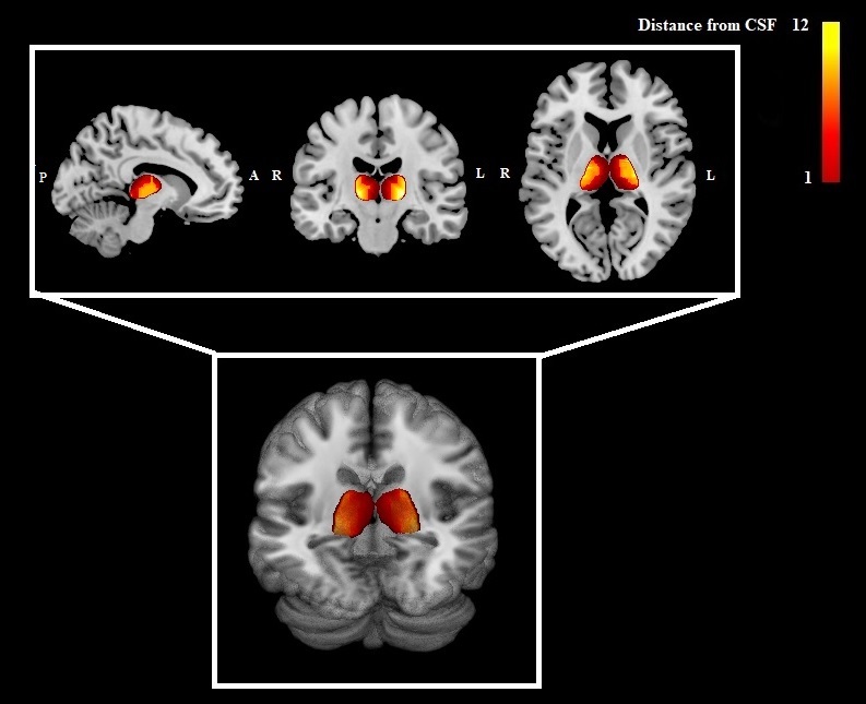
Figure 1. The Figure shows a map of the thalamus
of a MS patients color-coded according to the geodesic distance from the CSF (in
voxels) in the sagittal (on the left), coronal (in the middle) and axial (on
the right) plans in the first row. In the second row, a 3D rendering of the
same geodesic distance map of the thalamus. P=posterior; A=anterior; R=right;
L=left.
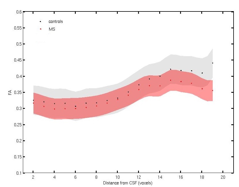
Figure 2.
In the graph, the mean (dots) and standard deviation (shadow)
FA values within the thalamus for healthy controls (black) and MS patients
(red) according to the distance from CSF (in voxels).
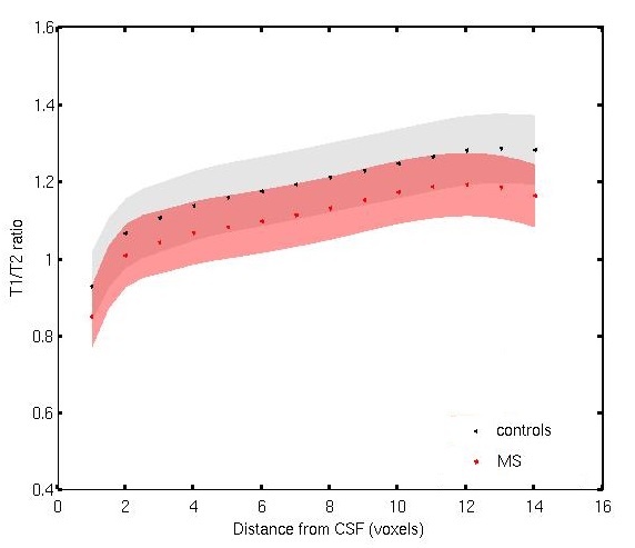
Figure 3. In the graph, the mean (dots)
and standard deviation (shadow) T1/T2-weighted
ratio values within the thalamus for
healthy controls (black) and MS patients (red) according to the distance from
CSF (in voxels).