3140
Thalamic energy dysregulation drives microstructural changes of thalamo-cortical projections in multiple sclerosis1Brain and Spinal Cord Institute (ICM), Paris, France, 2Department of Primary Care and Public Health, School of Public Health, Imperial College of London, London, United Kingdom, 3Department of Public Health, University “Federico II” of Naples, Naples, Italy, 4Neuroimaging Research Centre - ICM, Paris, France, 5Hopital De La Pitie Salpetriere, Paris, France, 6APHP, St Antoine Hospital, Neurology Department, Paris, France
Synopsis
Our objective was to investigate whether thalamic energy alterations in multiple sclerosis (MS) are associated with microstructural degeneration of thalamo-cortical tracts. In 17 patients and 13 healthy controls (HCs), the apparent diffusion coefficient of creatine-phosphocreatine ADC(tCr) in the thalami, reflecting energy dysregulation, was evaluated with diffusion-weighted magnetic resonance spectroscopy. Integrity of thalamo-cortical and non-thalamic tracts was evaluated by measuring mean diffusivity (MD) with standard diffusion weighted imaging. In patients but not in HCs, lower thalamic ADC(tCr) was associated with higher MD of thalamo-cortical tracts only, suggesting that thalamic energy dysfunction may induce the selective anterograde degeneration of thalamo-cortical networks.
INTRODUCTION
Neuronal energy dysfunction in multiple sclerosis (MS) has been proposed as a key driver of axonal degeneration. Diffusion-weighted 1H magnetic resonance spectroscopy (DW-MRS) allows to measure the diffusivity of creatine-phosphocreatine (tCr), a tandem of metabolites involved in adenosine triphosphate (ATP) production. DW-MRS combines the metabolite-specific information characteristic of MRS with the sensitivity to tissue microstructure characteristic of DW imaging. Using DW-MRS, we recently described in the thalamus of patients with MS a reduction of tCr diffusivity, reflecting energy dysregulation of thalamic neuronal and glial cells1. Energy failure at the thalamic level may result in a disruption of thalamo-cortical networks2. To test this hypothesis, we explored in vivo whether thalamic energy dysregulation is associated with microstructural degeneration of thalamo-cortical projections in MS.METHODS
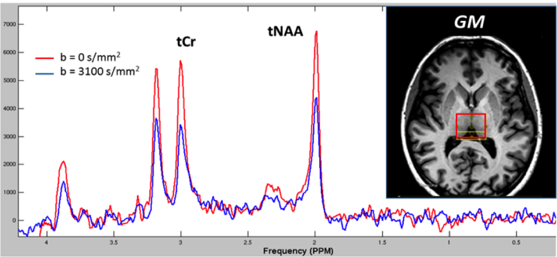
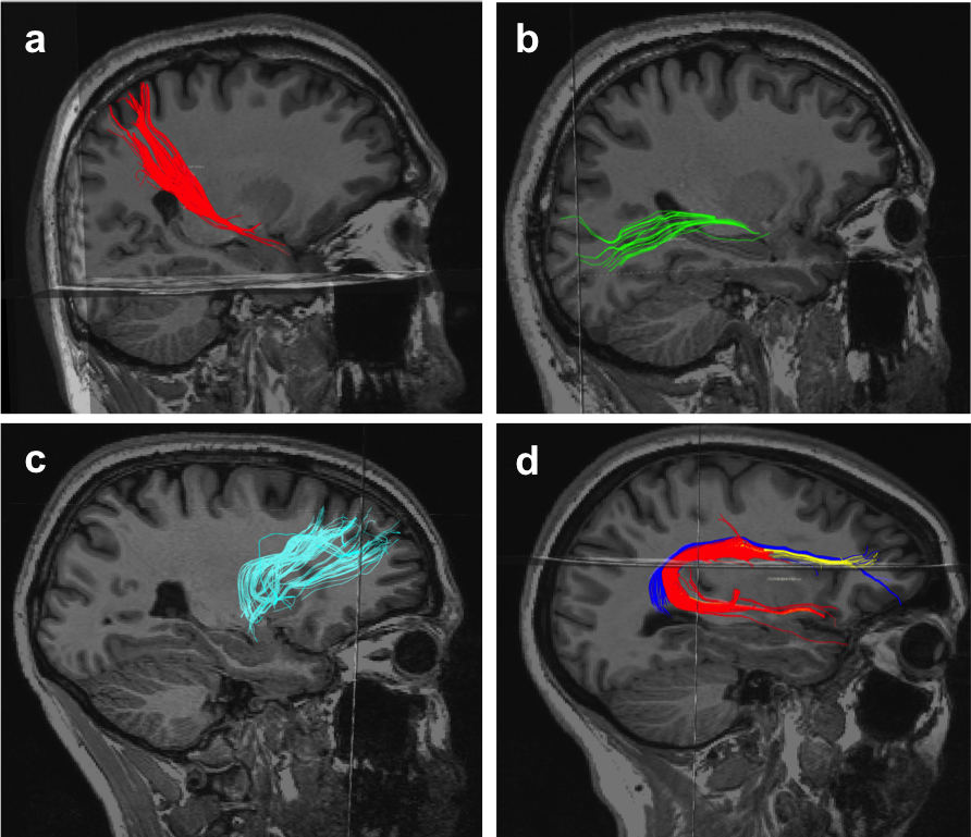
Seventeen patients with MS and thirteen healthy controls (HCs) were scanned using a 3T Siemens MAGNETOM Trio MRI scanner equipped with a 32-channel head coil. The imaging protocol included T1- and T2-weighted sequences. White matter lesion masks were generated based on T2-weighted images. Diffusion-weighted spectra were acquired using a single-voxel, PRESS sequencecombined with bipolar diffusion gradients. A VOI of 23(AP) × 30(LR) × 18(FH) mm3 was positioned in the bilateral thalami (Figure 1). Sequence parameters were TR=3 cardiac cycles, TE=120ms, spectral width = 2 kHz, number of points = 1024. Diffusion weighting was applied in one direction using a diffusion time of 60ms, gradient duration of 30ms, and a b-value of 3100 s/mm2. For each condition, including a non diffusion-weighted acquisition, 32 spectra were acquired. Water suppression was applied using a frequency selective excitation pulse followed by a dephasing gradient before metabolite excitation. Non-water suppressed spectra were acquired for eddy current corrections. Phase and frequency drift corrections were performed for individual spectra before summation in MATLAB. Quantification of spectral data was performed with LCModel3. The apparent diffusion coefficient of tCr, ADC(tCr), was obtained as the slope of the logarithm of the signal decay induced by diffusion weighting. DWI sequence included 50 DW directions with b=1000 s/mm2 and 1 image at b=0, with the following parameters: TR=7900ms, TE=87ms, Flip angle=90°, and voxel size=2 mm3. Images were preprocessed in ExploreDTI, including motion, eddy current and EPI distortion corrections, followed by the calculation of the diffusion tensor (DT) and maps of mean diffusivity (MD). Whole brain tractography was calculated using constrained spherical deconvolution with a maximum angle threshold of 30°. Thalamic bilateral projections to the somatosensory cortex (SS) and to the dorsolateral prefrontal cortex (DLPC), and the optic radiations (OR) (all white matter tracts connected with the thalamus), were manually drawn in TrackVis, along with the bilateral arcuate fasciculus, which was selected as control tract not connected with the thalamus (Figure 2a-d). Average bilateral mean diffusivity (MD) was extracted for each tract pair. The association between thalamic ADC(tCr) and MD of tracts was assessed in patients and HCs using linear mixed-effect regression models, adjusting for disease duration, gender, and (in patients only) tract-specific lesion load, calculated by overlapping lesion masks with the selected tracts in TrackVis.RESULTS
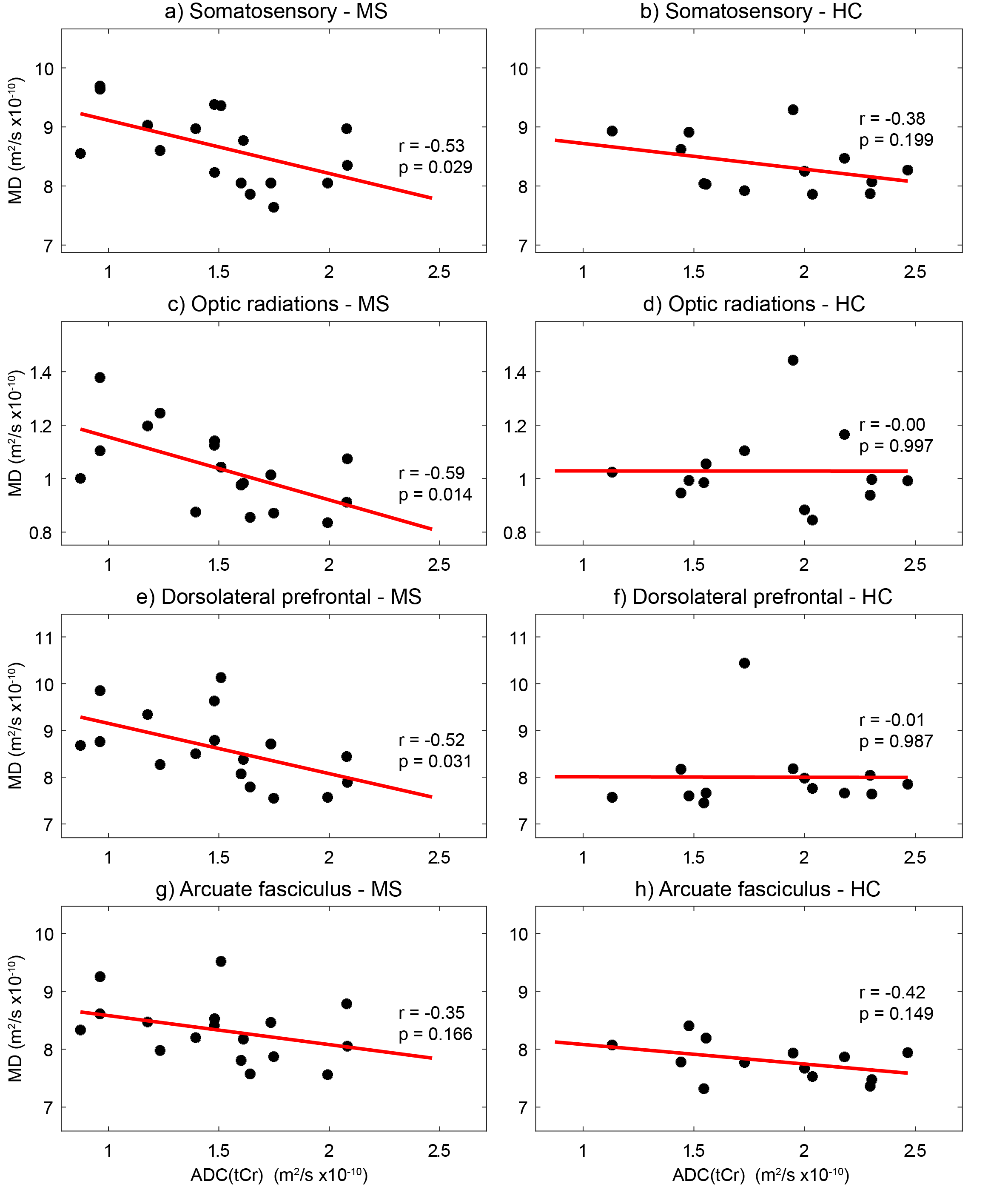
In patients with MS, lower thalamic ADC(tCr), reflecting energy dysregulation, was significantly associated with higher MD, reflecting microstructural changes, of pooled thalamo-cortical tracts, after adjusting for disease duration, gender and tract-specific lesion load (p=0.009), while no correlation was found in HCs (p=0.721). Unadjusted tract-by-tract analysis confirmed this association in patients (SS: p=0.029; DLPF: p=0.031; OR: p=0.014, respectively, Figure 3). Thalamic ADC(tCr) was not associated with the MD of the control tract (arcuate fasciculus, p=0.166).DISCUSSION
We demonstrated a significant correlation between energy dysregulation in the thalami and microstructural changes of thalamo-cortical projections in patients with MS. This correlation is disease-specific, as it was not found in HCs, and strictly limited to the tracts connected to the thalamus, as it was not shown in the control tract. A possible interpretation of these results is that the energy distress of neuronal bodies at the thalamic level spreads towards the axons of connected tracts, ultimately inducing their microstructural alteration. Another key finding is that the association between thalamic energy dysregulation and microstructural changes of connected tracts is independent of tract-specific lesions. This finding suggests that reduced thalamic energy levels may per se trigger neuroaxonal degeneration, independently of the damage resulting from demyelination, as previously suggested4.CONCLUSIONS
Energy dysregulation of thalamic cells was associated with microstructural changes of thalamo-cortical projections in patients with MS. Identifying the key steps linking the early phase of reduced energy supply in this hub with the progressive anterograde degeneration of connected fibers will be crucial to develop strategies to revert this process and, ultimately, to prevent neurodegeneration.Acknowledgements
No acknowledgement found.References
1. Bodini B, et al. Dysregulation of energy metabolism in multiple sclerosis measured in vivo with diffusion-weighted spectroscopy. Mult Scler 2018;24:313-321.
2. Tewarie P, et al. Functional brain networks: Linking thalamic atrophy to clinical disability in multiple sclerosis, a multimodal fMRI and MEG study. Hum Brain Mapp 2015;36:603–618.
3. Provencher SW. Automatic quantitation of localized in vivo 1H spectra with LCModel. NMR Biomed 2001;14:260–264.
4. Tallantyre EC et al, Clinico-pathological evidence that axonal loss underlies disability in progressive multiple sclerosis. Mult Scler 2010;16:406–411.
Figures