3137
Functional connectivity changes of nucleus accumbens subdivisions in left mTLE patients1Department of Radiology, The second Xiangya Hospital, Central South University, Changsha, China, 2Department of Medical Imaging Center, Nanfang Hospital, Southern Medical University, Guangzhou, China
Synopsis
Within the nucleus accumbens(NAC) there is a clear distinction between the shell and core portions. Growing evidences have supported that the NAC, especially its shell portion has been involved in epileptogenesis. However relevant studies on vivo human brains are quite limited. in this study, we investigated Left mTLE related function connectivity changes of NAC subregions . Our result indicated an decrease in FC between left shell and right frontal area and an increase FC between right shell and left temporal area. But no significant FC changes appear on core, which suggest that shell portion play important roles on mTLE.
Introduction
The nucleus accumbens (NAC) of human brain is an integral and specialized part of the ventral striatum[1]. Abnormalities within this nucleus have been proposed to underlie numerous psychiatric disorders, including schizophrenia, drug addiction, depression, obsessive/compulsive disorder and epilepsy. Therefore, it has been an important target for deep brain stimulation (DBS) and novel biological therapies of these diseases[2, 3]. However, our understanding of how pathophysiology within the NAC contribute to symptoms of these diseases is relatively infancy[4]. Within the NAC there is a clear distinction between the shell and core portions defined by their distinct cytoarchitectonics and connectivity patterns. Growing evidences have supported that the NAC, especially its shell portion has been involved in epileptogenesis. However relevant studies on vivo human brains are quite limited. Our previous study parcellated NAC into distinct subregion in mesial temporal lobe epilepsy (mTLE) patients using DTI connectivity-based parcellation[5], based on which, in this study, we investigated Left mTLE related function connectivity (FC) changes between NAC subregions and whole brain using functional magnetic resonance imaging (fMRI).Method
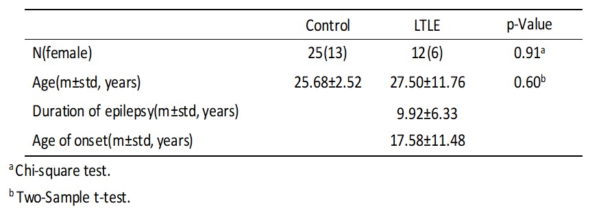
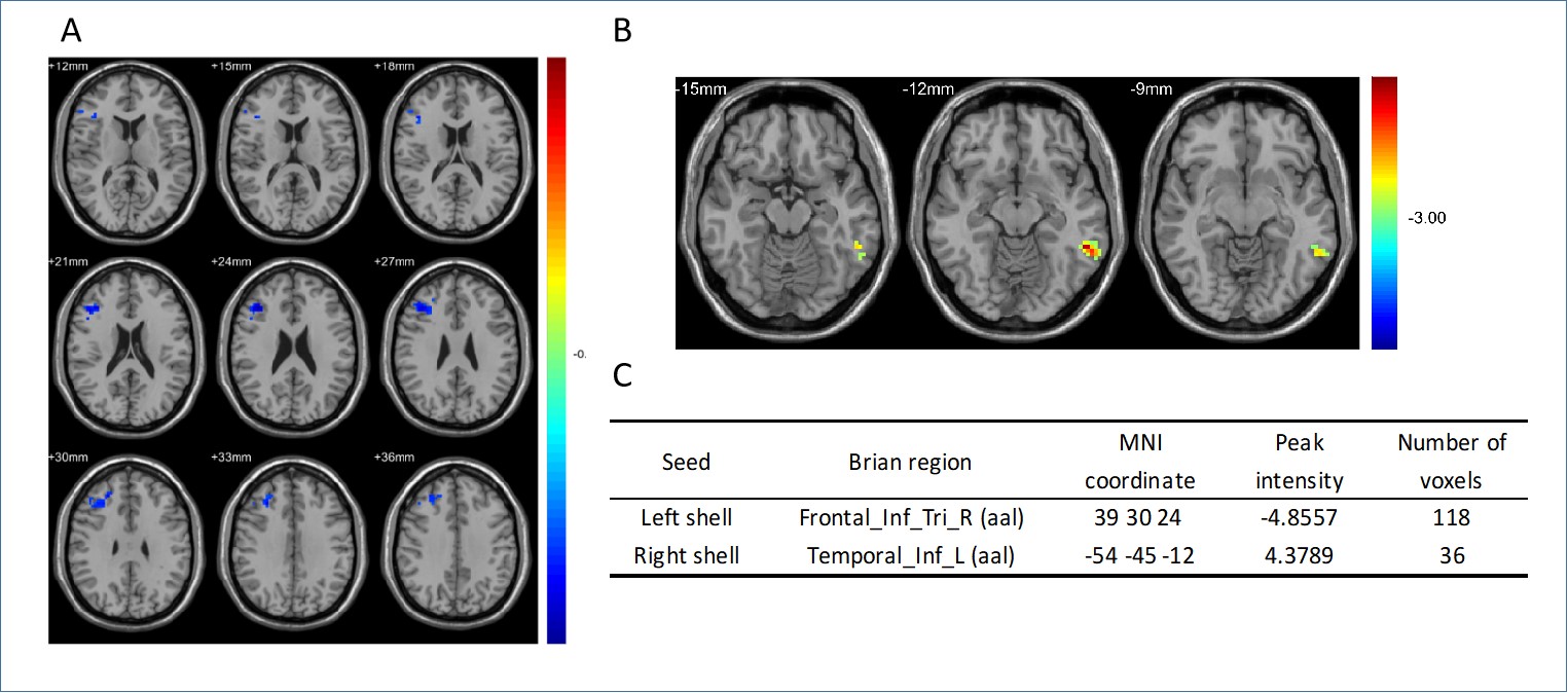
Subjects and data acquisition: Our study was conducted on 17 left mTLE patients and 25 healthy controls. All data were acquired on a 3.0T MR scanner (Philips Achieva TX) with 8-channel head coil. A fast field echo-planar imaging (FFE-EPI) protocol was acquired for rs-fMRI with TR/TE=2000/35 ms, voxel size=3×3×3 mm3, FOV= 240×240mm2, 33 slices and 300 repeated volumes. Data analysis: The left core and shell subregions which derived in our previous study were overlapped in standard space to create group seed ROIs. The resting-state fMRI data were preprocessed and then Pearson correlation coefficients between time series of each seed ROI and whole brain were computed. Two sample T-test was performed to identify the effect of the disease on each seed’s connectivity separately. The statistical result was then corrected for multiple comparisons using AlphaSim. A combination threshold of voxels' P<0.005 and clusters’ P<0.05 was considered significant.Results
Patient group and healthy control group were comparable in age and gender as showed in Table 1. Compared to healthy control, neither left or right core show significant FC changes in left mTLE group. On the contrary, the left shell shows decreased FC with a cluster located in right Frontal Lobe (Figure 1 A, C). The right shell portion shows increased FC with a cluster mainly located in left Temporal Lobe (Figure 1 B, C).Discussion and Conclusions
This is the first study to investigate the functional connectivity changes of NAC sudivisions of epilepsy in vivo human brain. It has been proved by various study that input from the temporal and frontal lobes display distinct topographical organization throughout NAC subregions[6, 7]. A temporal lobe seizure may spread to many brain areas directly and in directly connected to the temporal lobe, which include limbic and frontal brain structures[8, 9]. Our result indicated an decrease in FC between left shell and right frontal area and an increase FC between right shell and left temporal area, which suggests that NAC, especially its shell portion play important roles on mTLE. Moreover, our result shows that the left mTLE related changes on NAC are mainly on shell portion rather than core, which is in accordance with our both our assumption and previous result from DTI data[5] that neuronal degeneration and changed related to seizure mainly exists in shell. However, the detailed mechanism behind the FC changes of NAC shell related to mTLE need to be future investigated.Acknowledgements
No acknowledgement found.References
[1]. Heimer, L., et al., Specificity in the projection patterns of accumbal core and shell in the rat. Neuroscience, 1991. 41(1): p. 89-125.
[2]. Francis, T.C., et al., Nucleus Accumbens Medium Spiny Neuron Subtypes Mediate Depression-Related Outcomes to Social Defeat Stress. Biological Psychiatry, 2015. 77(3): p. 212-222.
[3]. Salgado, S. and M.G. Kaplitt, The Nucleus Accumbens: A Comprehensive Review. Stereotactic & Functional Neurosurgery, 2015. 93(2): p. 75-93.
[4]. Floresco, S.B., The nucleus accumbens: an interface between cognition, emotion, and action. Annual review of psychology, 2015. 66: p. 25-52.
[5]. Zhao, X., et al., Connectivity-based parcellation of the nucleus accumbens into core and shell portions for stereotactic target localization and alterations in each NAc subdivision in mTLE patients. Human Brain Mapping, 2017. 39(3).
[6]. Groenewegen, H.J., et al., Organization of the projections from the subiculum to the ventral striatum in the rat. A study using anterograde transport of Phaseolus vulgaris leucoagglutinin. Neuroscience, 1987. 23(1): p. 103-120.
[7]. Brog, J.S., et al., The patterns of afferent innervation of the core and shell in the “Accumbens” part of the rat ventral striatum: Immunohistochemical detection of retrogradely transported fluoro-gold. Journal of Comparative Neurology, 1993. 338(2): p. 255-278.
[8]. Bertram, E.H., et al., Functional anatomy of limbic epilepsy: a proposal for central synchronization of a diffusely hyperexcitable network. Epilepsy Research, 1998. 32(1-2): p. 194-205.
[9]. Bartolomei, F., et al., Pre-ictal synchronicity in limbic networks of mesial temporal lobe epilepsy. Epilepsy Research, 2004. 61(1): p. 89-104.
Figures