3133
Surfaces Area and Cortical Volume Development of Infant Transverse Temporal Cortex Influenced by Preterm Birth1Department of Radiology, the First Affiliated Hospital, Xi’an Jiaotong University, Xi'an, China, 2Department of Radiology and BRIC, University of North Carolina at Chapel Hill, Chapel Hill, NC, United States, 3MR Research China, GE Healthcare, Beijing, China
Synopsis
Development of transverse temporal cortex is essential for speech perception in infants. However, the early morphological development of this cortex has not been fully understood. Additionally, influences of preterm birth on cortical development remain to be investigated. This study assessed cortical development of transverse temporal cortex in preterm and term infants based on surface reconstruction. We found that surface area and cortical volume of transverse temporal cortex underwent rapid changes. Term infants held higher surface area, cortical volume, and asymmetry than preterm infants. These results suggest that preterm birth influences the asymmetry and developmental trajectory of transverse temporal cortex.
INTRODUCTION
Transverse temporal cortex plays a crucial role in speech perception 1. Cortical regions with left-lateralization could already be evoked by speech in infants, similar to those of adults 2. This may be related to the early development of transverse temporal cortex 3. However, the development of this cortex in infants has not been fully understood, because of the challenges in the tissue segmentation and surface reconstruction. Additionally, the influence of preterm birth on cortical development remains to be investigated. Recently, effective methods have been proposed for processing the structural MRI 4, 5, which make it possible to answer the questions. In this study, we tried to assess the structural development of transverse temporal cortex in preterm and term infants based on cortical surface reconstruction.METHODS
This study was approved by the local institutional review board. Informed written consents were obtained from parents of the infants. Infants who were confirmed or suspected to have congenital malformations of central nervous system, congenital infections, metabolic disorders, abnormal appearances in MRI were excluded.
Three-dimensional fast spoiled gradient-recalled echo T1WI was performed on a 3T MRI scanner (Signa HDxt; GE Healthcare, USA) with an 8-channel head coil. Repetition time = 10.468 ms; Echo time = 4.764 ms; Inversion time: 400 ms; Field of view = 240 × 240 mm2; Acquisition matrix = 240 × 240; Reconstruction matrix = 256 × 256; Slice thickness = 1 mm.
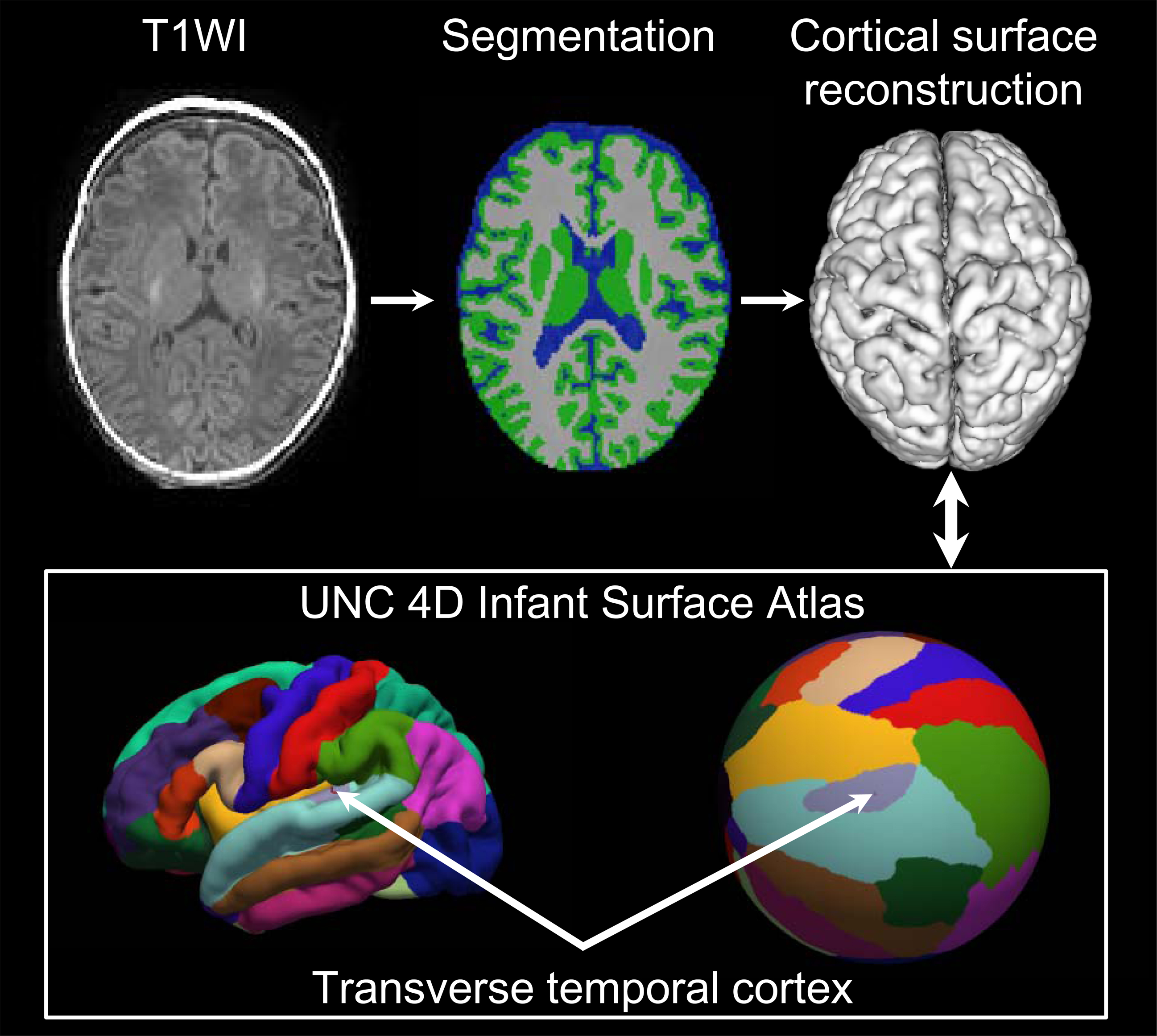
T1WI images were processed by using the UNC Infant Pipeline (Figure 1) 5. Firstly, segmentation of brain tissues was performed by using a deep learning method 4. Secondly, reconstruction of the topologically-correct and accurate cortical surfaces were performed. Thirdly, Cortical surfaces were mapped on to a spherical surface 6, aligned to the UNC 4D Infant Surface Atlas (https://www.nitrc.org/projects/infantsurfatlas). According to the atlas, left and right transverse temporal cortices were selected as regions of interest. Finally, surface area, cortical thickness, and cortical volume in transverse temporal cortices were computed 7, 8. Asymmetry index was calculated by the following equation: Asymmetry index = (Right−Left) / 0.5*(Right + Left) 9.
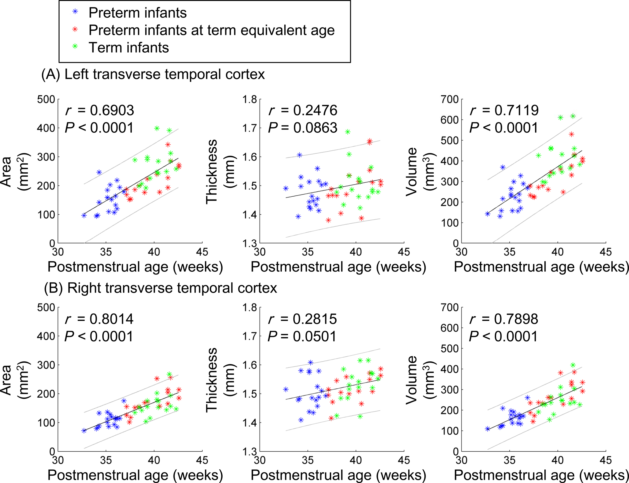
Inter-group differences in gestational age, postnatal ages at MRI, postmenstrual age (PMA), and regional values of metrics were evaluated using Mann-Whitney U test. Correlations between surface area, cortical thickness, volume and the postmenstrual age were performed using Spearman partial correlation after controlling the preterm/term factor. Chi-Square test was performed to evaluate gender ratio differences across groups. Tests were considered significant at P<0.05.
RESULTS
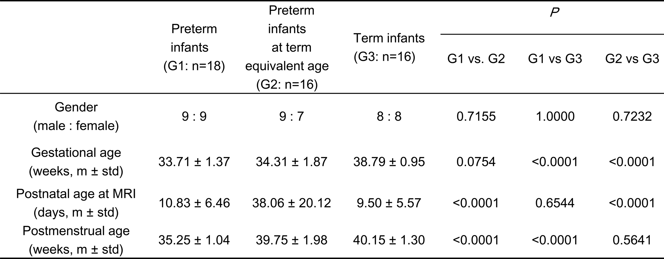
A total of 50 infants were enrolled, including 34 preterm and 16 term infants. Out of the preterm infants, 16 subjects were investigated at the term equivalent age (Table 1).
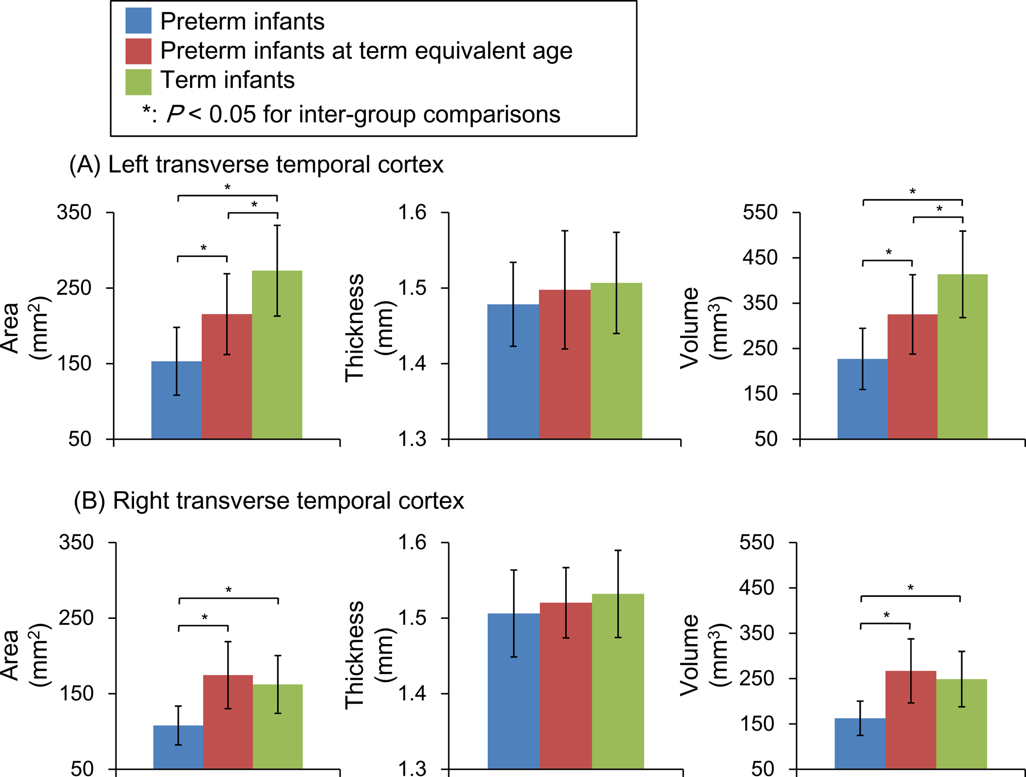
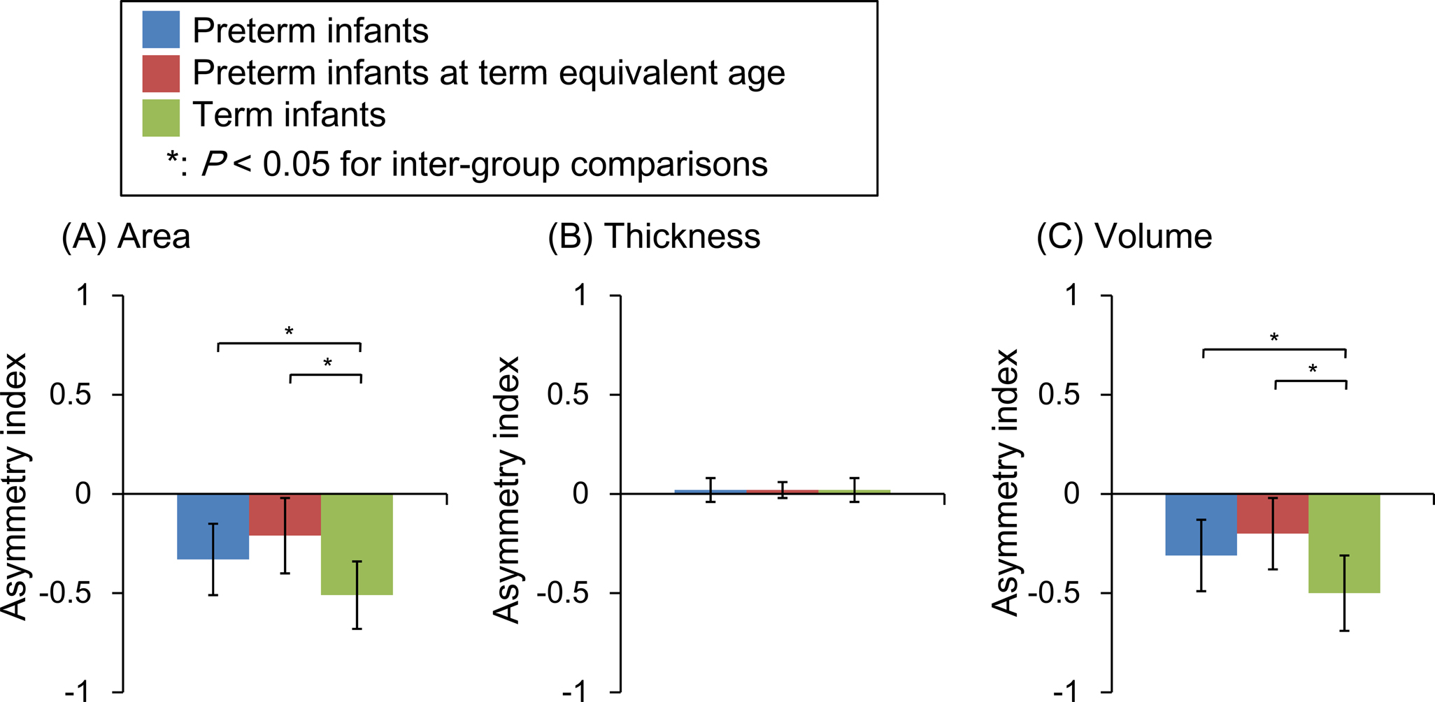
PMA related changes were observed in surface area and cortical volume (Figure 2), while no significant correlation between cortical thickness and PMA could be found. This developmental change pattern could also be found through inter-group comparisons (Figure 3). Compared to preterm infants at term equivalent age, term infants held bigger surface area and cortical volume in the left transverse temporal cortex, while no significant differences were found in the right (Figure 3).
Obvious asymmetry could be found in surface area and cortical volume, while the asymmetry index was low in cortical thickness (Figure 4). Higher asymmetry of surface area and cortical volume could be observed in term infants than those in preterm infants.
DISCUSSION
This study found that infant transverse temporal cortex underwent rapid structural changes. Significant age-related changes in surface area and cortical volume but no significant changes in thickness reflected the rapid folding during early development 10. Additionally, preterm infants could catch up term infants in surface area and cortical volume in the right transverse temporal cortex. However, there are still significant differences in the left hemisphere, even at term equivalent age. This reflects asymmetric structural development in cortex, which provides structural evidence for left-lateralization during the speech perception in infants 2.
Though the asymmetry was found in both preterm and term infants, term infants held higher asymmetry. Early exposure to extrauterine environment would influence the cortical folding 11. High rate of language learning difficulties in preterm population 12 may be associated with the early poor maturation revealed in this work.
CONCLUSION
Left and right transverse temporal cortices on infants undergo asymmetric development in surface area and cortical volume. And preterm birth influences the asymmetry and developmental trajectory of transverse temporal cortex.Acknowledgements
Xianjun Li, Jing Xia, Jian Chen contributed equally to this work. Please address correspondence to Gang Li, gang_li@med.unc.edu; Dinggang Shen, e-mail: dinggang_shen@med.unc.edu; and Jian Yang, e-mail: cjr.yangjian@vip.163.com. This study was supported by the National Key Research and Development Program of China (2016YFC0100300), National Natural Science Foundation of China (81471631, 81771810 and 81171317), the 2011 New Century Excellent Talent Support Plan of the Ministry of Education, China (NCET-11-0438), the Fundamental Research Funds for the Central Universities (xjj2018265), the Fundamental Research Funds of the First Affiliated Hospital of Xi'an Jiaotong University (2017QN-09).References
1. Hickok G, Poeppel D. The cortical organization of speech processing. Nature Reviews Neuroscience 2007;8:393-402
2. Dehaenelambertz G, Dehaene S, Hertzpannier L. Functional neuroimaging of speech perception in infants. Science 2002;298:2013-2015
3. Warrier C, Wong P, Penhune V, et al. Relating structure to function: Heschl's gyrus and acoustic processing. Journal of Neuroscience 2009;29:61-69
4. Wang L, Li G, Adeli E, et al. Anatomy‐guided joint tissue segmentation and topological correction for 6‐month infant brain MRI with risk of autism. Human Brain Mapping 2018;39:2609-2623
5. Li G, Wang L, Feng S, et al. Construction of 4D high-definition cortical surface atlases of infants: Methods and applications. Medical Image Analysis 2015;25:22-36
6. Yeo BTT, Sabuncu MR, Vercauteren T, et al. Spherical Demons: Fast Diffeomorphic Landmark-Free Surface Registration. IEEE Transactions on Medical Imaging 2010;29:650-668
7. Li G, Lin W, Gilmore JH, et al. Spatial Patterns, Longitudinal Development, and Hemispheric Asymmetries of Cortical Thickness in Infants from Birth to 2 Years of Age. Journal of Neuroscience 2015;35:9150-9162
8. Li G, Nie J, Wang L, et al. Mapping region-specific longitudinal cortical surface expansion from birth to 2 years of age. Cerebral Cortex 2013;23:2724-2733
9. Toga AW, Thompson PM. Mapping brain asymmetry. Nature Reviews Neuroscience 2003;4:37-48
10. Jha SC, Xia K, Ahn M, et al. Environmental Influences on Infant Cortical Thickness and Surface Area. Cerebral Cortex 2018
11. Lefèvre J, Germanaud D, Dubois J, et al. Are Developmental Trajectories of Cortical Folding Comparable Between Cross-sectional Datasets of Fetuses and Preterm Newborns? Cerebral Cortex 2015;26:99-100
12. Zambrana IM, Vollrath ME, Sengpiel V, et al. Preterm delivery and risk for early language delays: a sibling-control cohort study. International Journal of Epidemiology 2015;45:151-159
Figures