3132
Cortical FoldingPrint for Individual Identification During Dynamic Postnatal Development1Key Laboratory of Biomedical Engineering of Ministry of Education, Zhejiang University, Hangzhou, China, 2Department of Radiology and BRIC, University of North Carolina at Chapel Hill, Chapel Hill, NC, United States, 3Department of Psychiatry, University of North Carolina at Chapel Hill, Chapel Hill, NC, United States
Synopsis
Human cortical folding is highly convoluted and characterizes the inter-subject variability. Recent studies found that the adult brain cortex is unique for individual identification. However, little is known about whether the infant brain cortex, which develops dynamically in the first postnatal years, is reliable for individual identification. To this end, we proposed a novel morphological folding descriptor, called FoldingPrint, to perform the infant identification in a large longitudinal dataset with 472 infants. Successful identification results indicate the effectiveness of the proposed FoldingPrint. In addition, we found that the regions with high identification accuracy are mainly distributed in high-order association cortices.
Introduction
The human cortical folding is highly convoluted in shape, which characterizes the inter-subject variability. Current studies found that the structural (e.g., BrainPrint1) or functional (e.g., functional connectome fingerprinting2,3) features of the adult brain are unique and reliable for individual identification. However, these studies were all performed on adult brains, which have relatively subtle changes across different scans. In contrast, the infant identification is more challenging, since the infant cortex develops dynamically and regionally-heterogeneously in the first postnatal years. Thus, the dedicated shape descriptor needs to be stable during the early brain development. To this end, we propose a novel morphological folding descriptor based on multi-scale curvatures, called FoldingPrint, for infant identification. This study thus aims to address two essential neuroscience questions: 1) whether the developing infant cortical folding is unique for individual identification; 2) which cortical regions are more distinct and reliable for infant identification.Methods
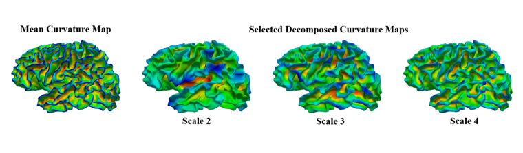
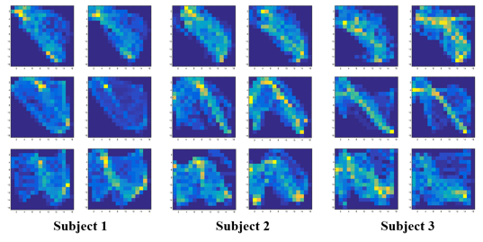
Longitudinal T1w and T2w MR images of 472 healthy infants at 0, 1, and 2 years of age, with totally 1,141 scans, were acquired on a 3T Siemens scanner. All MR images were processed via an infant-dedicated computational pipeline4. Each reconstructed cortical surface was aligned onto the age-matched template in the UNC 4D infant cortical surface atlas5 to obtain a parcellation map6 with 34 regions of interest (ROIs). We created FoldingPrint through the following steps: first, the mean curvature map of each inner cortical surface was decomposed into multi-scales using over-complete spherical wavelets7, thus obtaining a comprehensive characterization of cortical folding. Second, in each ROI, the three most informative curvature scales (scales 2, 3, and 4 as shown in Fig. 1) were selected to construct a high-dimensional folding descriptor, i.e., FoldingPrint, based on their joint probability density as shown in Fig. 2. As we can see, the feature patterns on the 2D projections of the FoldingPrint are variable across different subjects, but stable across different ages of the same subject. Thus, the proposed descriptor can be used for characterizing the developing cortical folding for infant identification. Third, given a new scan to be identified, its FoldingPrint was compared to that of each scan in the existing database in each ROI. Herein, the Chi-squared distance was adopted to measure the similarity of each ROI’s FoldingPrint between different subjects. In each ROI, the subject with the highest similarity was regarded as the potentially-identified subject. In total, 68 results were obtained from all ROIs in both hemispheres. Finally, through an ROI-based majority voting scheme, the potentially-identified subject with the highest frequency was considered as the finally-identified subject. In addition, to explore which regions are more reliable for individual identification, we further computed the ROI-specific identification accuracy maps.Results
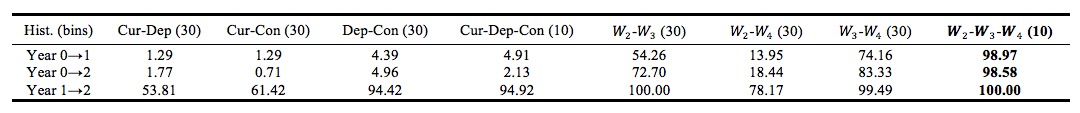
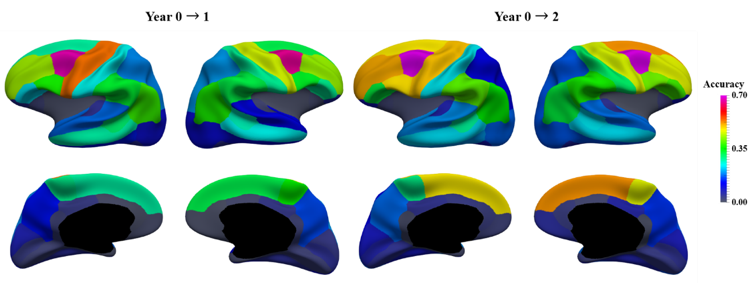
The experiments were carried out in 3 tasks: 1) using the scans at birth to identify their corresponding scans at year 1; 2) using the scans at birth to identify their corresponding scans at year 2; 3) using the scans at year 1 to identify their corresponding scans at year 2. Since cortical folding develops more rapidly in the first year than the second year, the first two tasks are much more challenging than the third one. Table 1 reports the identification accuracies of different high-dimensional feature combinations. The results of the proposed FoldingPrint with multi-scale curvatures W2-W3-W4 (10) are in bold. As we can see, compared to other different 2D combinations of decomposed curvatures, the proposed descriptor obtains the best performance on all three tasks, i.e., 98.97%, 98.58%, and 100.00%, respectively. Besides, from the results of high-dimensional feature combinations of the traditional cortical folding features, i.e., mean curvature, average convexity, and sulcal depth, we found that they still failed to show comparable performance as the proposed FoldingPrint, especially in the first two tasks. In addition, the maps of the region-specific accuracies in infant identification are displayed in Fig. 3. We found that the regions with high identification capability are mainly distributed in the high-order association cortices, especially in the prefrontal cortex.Discussion and Conclusion
We proposed a novel morphological descriptor based on multi-scale decomposed curvatures, i.e., FoldingPrint, for infant identification. The proposed descriptor is capable of comprehensively characterizing the invariance of developing cortical folding and achieves promising identification accuracy. In addition, we found that the high-order association cortices, especially the prefrontal cortex, encode more individual variability, thus presenting high identification capability. Besides, the patterns of the ROIs’ identification capability are region-specific and time-consistent. Future work will include identifying the twins and exploring their individual variability patterns for better understanding the genetic and environmental influences on the individual variability of cortical folding.Acknowledgements
This work was supported in part by NIH grants (MH100217, MH108914, MH107815, MH110274, MH070890, MH064065, HD053000, MH109773, MH116225 and MH117943).References
1. Wachinger C, Golland P, Kremen W, et al. BrainPrint: a discriminative characterization of brain morphology. Neuroimage. 2015;109(1): 232-248.
2. Finn E S, Shen X, Scheinost D, et al. Functional connectome fingerprinting: identifying individuals using patterns of brain connectivity. Nat Neurosci. 2015;18(11):1664-1670.
3. Liu J, Liao X, Xia M, et al. Chronnectome fingerprinting: identifying individuals and predicting higher cognitive functions using dynamic brain connectivity patterns. Hum Brain Mapp. 2018;39(2):902-915.
4. Li G, Wang L, Shi F, et al. Construction of 4D high-definition cortical surface atlases of infants: Methods and applications. Medical image analysis. 2015; 25(1):22-36.
5. https://www.nitrc.org/projects/infantsurfatlas/.
6. Desikan R S, Ségonne F, Fischl B, et al. An automated labeling system for subdividing the human cerebral cortex on MRI scans into gyral based regions of interest. Neuroimage. 2006;31(3):968-980.
7. Yeo B T T, Yu P, Grant P E, et al. Shape analysis with overcomplete spherical wavelets. MICCAI. 2008:468-476.
Figures