3126
Cortical and subcortical networks in frontal lobe epilepsy with generalized tonic clonic seizures1jinling hospital, nanjing, China
Synopsis
Cortico-subcortical networks are considered core pathologic substrates for frontal lobe epilepsy with generalized tonic clonic seizures; however, the mechanism is still unknown. This study aims to identify the changes of cortico-subcortical networks by resting-state functional connectivity. 60 patients with frontal lobe epilepsy and healthy controls were enrolled. Bilateral hemispheres were divided into 5 nonoverlapping cortical lobes. Functional connectivity between each cortical lobe and the subcortical regions were calculated, and functional connectivity strength was used to evaluate the interconnectivity. Our results indicate that the decrease connection between prefrontal cortex and subcortical structures suggests it maybe the epicenters of frontal lobe epilepsy.
INTRODUCTION
Epilepsy is a kind of disorder with abnormal brain network1. As the second highest type of localization-related epilepsies,frontal lobe epilepsy accounts for 20 to 30 percent among all partial epilepsies 2,3. Its transmission is realized by complex cortical and subcortical structure and functional network4. Thalamus, cerebellum and basal ganglia play an important role in the transmission of epilepsy, especially thalamus is considered as an important node of comprehensive seizure symptoms5-8. Patients with frontal lobe epilepsy with generalized tonic clonic seizures are considered to be a kind of focal epilepsy originating from the frontal cortex and spreading through the cortico-subcortical network, but the mechanism of its transmission is still unclear9-11. Therefore, this study intends to observe the changes of cortico-subcortical networks in patients with frontal lobe epilepsy with generalized tonic clonic seizures by resting functional magnetic resonance imaging in order to understanding the pathophysiology of this disorder.
METHODS
In this study, 60 patients with frontal lobe epilepsy and 60 age-and gender-matched healthy patients were enrolled. The data were preprocessed and analyzed by DPARSF and SPM software. The preprocessing included slice timing, head motion correction, normalize, smoothing, filtering and regression. According to previous research12,13, we divided bilateral hemispheres into 5 nonoverlapping cortical lobes based on anatomic templates: prefrontal cortex, motor/premotor cortex, somatosensory cortex, parietal/occipital cortex, and temporal cortex. Subcortical structures including thalamus, basal ganglia and cerebellum were defined by AAL template. Functional connectivity was conducted to identify the corticol and subcorticol networks. We applied the winner-take-all approach to compute the functional connectivity strength. Compare the connectivity between each cortical ROIs and the subcorticol structures among groups by using SPM software.RESULTS
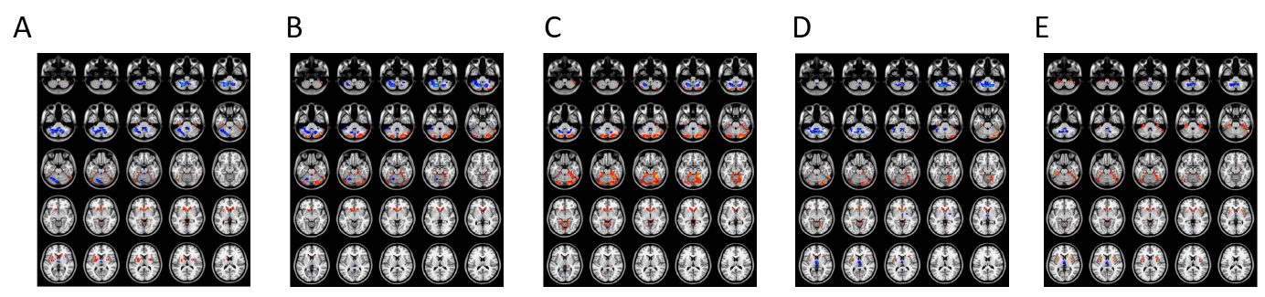
Compared with controls, the junction number between prefrontal cortex and subcortical structures decreased while temporal cortex, motor cortex, somatosensory cortex increased.In maps of connectivity strength, the connectivity between prefontal cortex and subcortical cortex were increase in the basal ganglia, and decrease in the cerebellum and thalamus. The connection between motor/premotor cortex and subcortical cortex were increase in the basal ganglia, decrease in the thalamus and shows both increase and decrease in the cerebellum. The connection between somatosensory cortex and subcortical cortex were increase in the basal ganglia and thalamus and shows both increase and decrease in the cerebellum. The connection between parietal/occipital cortex and subcortical cortex were increase in the basal ganglia, decrease in the thalamus and shows both increase and decrease in the cerebellum. The connection between temporal cortex and subcortical cortex were increase in the basal ganglia, decrease in the thalamus and shows both increase and decrease in the cerebellum.DISCUSSION
Our results indicate that frontal lobe epilepsy patients with generalized tonic clonic seizures is associated with the decrease connection between prefrontal cortex and subcortical structures which suggests that it maybe the epicenters of frontal lobe epilepsy.CONCLUSIONS
Since the basic pathophysiology of epilepsy is still not fully understood, our finding of the abnormality of the cortical and subcortical connection may provide insight into the pathophysiology of frontal lobe epilepsy patients with generalized tonic clonic seizures.Acknowledgements
We thank the patients and volunteers for participating in this study. We also thank the anonymous reviewers for their constructive suggestions to improve this work.References
1. Yuen A W C , Keezer M R , Sander J W . Epilepsy is a neurological and a systemic disorder[J]. Epilepsy & Behavior E & B, 2017, 78:57.
2. Cao X., Qian Z., Xu Q., Shen J., Zhang Z., Lu G. Altered intrinsic connectivity networks in frontal lobe epilepsy: a resting-state fMRI study. Comput Math Methods Med. 2014: 864979.
3. Woodward K E , Gaxiolavaldez I , Goodyear B G , et al. Frontal Lobe Epilepsy Alters Functional Connections Within the Brain's Motor Network: A Resting-State fMRI Study[J]. Brain Connectivity, 2014, 4(2):91-9.
4. Blumenfeld H, Varghese G I, Purcaro M J, et al. Cortical and subcortical networks in human secondarily generalized tonic–clonic seizures[J]. Brain, 2009, 132(4):999-1012.
5. Moeller F, Siebner H R, Wolff S, et al. Changes in activity of striato–thalamo–cortical network precede generalized spike wave discharges[J]. Neuroimage, 2008, 39(4):1839-1849.
6. Bernhardt B C , Rozen D A , Worsley K J , et al. Thalamo–cortical network pathology in idiopathic generalized epilepsy: Insights from MRI-based morphometric correlation analysis[J]. Neuroimage, 2009, 46(2):373-381.
7. Yang L , Li H , Zhu L , et al. Localized shape abnormalities in the thalamus and pallidum are associated with secondarily generalized seizures in mesial temporal lobe epilepsy[J]. Epilepsy & Behavior, 2017, 70(Pt A).
8. Raethjen J , Muthuraman M . Cause or compensation? Complex changes in cerebello-thalamo-cortical networks in pathological action tremor.[J]. Brain, 2015, 138(10):2808-2810.
9. Dong L, Wang P, Peng R, Jiang S, Klugah-Brown B, Luo C, Yao D. Altered basal ganglia-cortical functional connections in frontal lobe epilepsy: a resting-state fMRI study. Epilepsy Res, 2016, 128:12–20.
10. Vaessen M J , Braakman H M H , Heerink J S , et al. Abnormal Modular Organization of Functional Networks in Cognitively Impaired Children with Frontal Lobe Epilepsy[J]. Cerebral Cortex, 2013, 23(8):1997-2006.
11. Braakman H M H , Vaessen M J , Jansen J F A , et al. Frontal lobe connectivity and cognitive impairment in pediatric frontal lobe epilepsy.[J]. Epilepsia, 2013, 54(3):446-454.
12. Zhang D , Snyder A Z , Fox M D , et al. Intrinsic Functional Relations Between Human Cerebral Cortex and Thalamus[J]. Journal of Neurophysiology, 2008, 100(4):1740.
13. Ji G J, Zhang Z, Xu Q, et al. Identifying Corticothalamic Network Epicenters in Patients with Idiopathic Generalized Epilepsy[J]. Ajnr American Journal of Neuroradiology, 2015, 36(8):1494.
Figures