3123
A comparison of high b-value and standard b-value diffusion weighted imaging for status epilepticus in pediatric patients1Department of Radiology, The First Affiliated Hospital of Xiamen University, Xiamen, China, 2Department of Nuclear Medicine and Minnan PET Center, The First Affiliated Hospital of Xiamen University, Xiamen, China
Synopsis
In this study series with status epilepticus (SE) pediatric patients, we investigated the utility diffusion weighted magnetic resonance imaging with a high b-value (b = 3000 s/mm2) compared with standard b-value (b = 1000 s/mm2) for acute ictal MRI changes in pediatric patients. High b-value diffusion weighted imaging (DWI) could be beneficial for detecting additional lesions and improving the contrast between lesions and normal tissue. Therefore, high b-value DWI may be a better noninvasive imaging method for exploration of the acute ictal MRI changes in pediatric patients with SE.
INTRODUCTION
Electroencephalography (EEG) is a standard examination for the diagnosis of status epilepticus but is not necessarily available in the emergency department of general hospitals without a medical center specializing in epilepsy. Some reports have described SE showing hyperintense areas on magnetic resonance standard b-value DWI. DWI imaging with higher b-values was better than standard b-value imaging for diagnosis of acute stroke[1], Cretzfeldt-Jakob disease[2], and showed better contrast between lesions and normal tissue in primary central nervous system lymphoma[3] and medulloblastoma[4] in our previous studies. However, high b-value DWI has not yet been evaluated in the acute ictal setting with respect to the alterations in brain tissue.METHODS
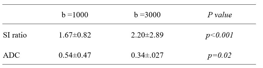
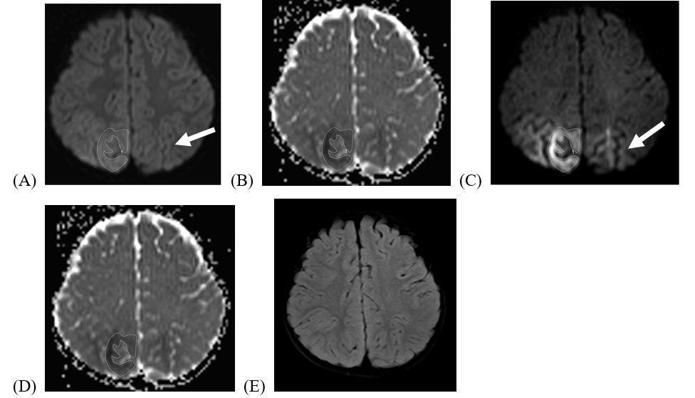
High and standard b-value (b = 3000 and 1000 s/mm2, respectively) DWI were acquired in 10 pediatric patients with SE. Two patients with no underlying disease as potential cause for the seizures (n= 2), four patients with fever (n= 4), two patients with mitochondrial encephalopathy lactic acidosis and stroke-like episode (MELAS) (n = 2) and two patients with acute encephalopathy with biphasic seizures and late reduced diffusion (AESD) (n =2) were included in this study. The diagnostic quality of brain lesion was compared between b = 1000 and b = 3000 s/mm2 DWI images by visual inspection. In addition, we attempted a quantitative assessment using apparent diffusion coefficient (ADC) value and an index of signal intensity ratio, defined as the mean SI at the affected lesion divided by the mean SI at the pons[5].RESULTS
Twenty lesions in ten patients were revealed. Fifteen of the lesions were hyperintense at DWI with high and standard b-values; high b-value images revealed five additional lesions (Fig 1). Lesions were also more conspicuous at high b-value DWI compared to standard b-value images. The mean SI ratio was significantly greater in the b =3000 s/mm2 images compared to the b =1000 s/mm2 images. Mean ADC values were lower in the all the subjects on b= 3000 s/mm2 images than that on b = 1000 s/mm2 images (Table1).DISCUSSION
The present report is the first to focus on the clinical efficacy of high b value DWI in cases of acute itcal phase of SE during childhood, along with comparison of standard b-value DWI and ADC. Higher b-value (b =3000 s/mm2) was benefical for detecting additional lesions and improving the contrast between lesions and normal tissue.CONCLUSIONS
We concluded that b =3000 s/mm2 DWI was superior to b = 1000 s/mm2 DWI in detecting abnormal lesions in the acute itcal phase of SE for pediatric patients.Acknowledgements
No acknowledgement found.References
1. Purroy F, Begue R, Quilez A, et al. Contribution of high b-value diffusion-weighted imaging in determination of brain ischemia in transient attack patients. J Neuroimaging 2013;23:33-38.
2. Hyare H, Thornton J, Stevens J et al. High b-value diffusion MR imaging and basal nuclei apparent diffusion-weighted MRI in the diagnosis of Creutzfeldt-Jakob disease. AJNR Am J Neuroradoil 2010;31:521-526.
3. Han H, Han C, Huang S, et al. Comparison of diffusion-weighted imaging between high and standard b-values for primary central nervous system lymphoma. Clin Radiol 2014; 69: 974-979.
4. Han C, Zhao L, Wu X, et al. A comparison of high b-value vs standard b-value diffusion weighted magnetic resonance imaging at 3.0T for Medulloblastoma. Br J Radiol 2015; 88:20150220.
5. Tsubouchi Y, Itamura S, Saito Y et al. Use of high b value diffusion-weighted magnetic resonance imaging in acute encephalopathy/encephalitis during childhood. Brain Dev 2108; 40:116-125.
Figures