3120
Community-informed connectomics of the thalamo-cortical system in idiopathic generalized epilepsy1The Affiliated Hospital of Nanjing University Medical School, Nanjing, China, 2McGill University, Montreal, QC, Canada
Synopsis
Idiopathic generalized epilepsy with tonic-clonic seizures (IGE-GTCS) has been associated to the thalamo-cortical circuitry. By quantifying the interplay between macroscale functional communities via resting-state fMRI (rs-fMRI) connectome analysis, we assessed the intrinsic organization of this network and its relation to drug-response. Compared to controls, IGE-GTCS showed a more constrained network embedding of the thalamus, while frontocentral neocortical regions expressed increased functional diversity. Findings remained significant after regressing out thalamic volume and cortical thickness, suggesting independence from structural alterations. We observed more marked network imbalances in drug-resistant compared to seizure-free patients. Our findings suggest a pathoconnectomic mechanism of IGE, centered on diverging changes in cortical and thalamic connectivity. More restricted thalamic connectivity could reflect the tendency to engage in recursive thalamo-cortical loops, which may contribute to hyper-excitability.
Introduction
Idiopathic generalized epilepsy with tonic-clonic seizures (IGE-GTCS) is a disabling epilepsy syndrome, with 30% drug-resistant patients. Electrophysiological and MRI studies suggest a key role of thalamo-cortical networks in its pathophysiology. MRI studies have mapped structural changes and atypical connectivity, but did not address how localized anomalies may translate into macroscale pathophysiology, particularly with respect to drug- response. Connectomics taps into whole-brain organization and decomposability into interacting communities. We studied the thalamo-cortical system in a prospective sample of IGE-GTCS and healthy controls using community-informed connectomics. Analysis was complemented with MRI morphometry, to determine functional perturbations above and beyond structural compromise. We furthermore examined associations between network anomalies and drug-response.
Methods
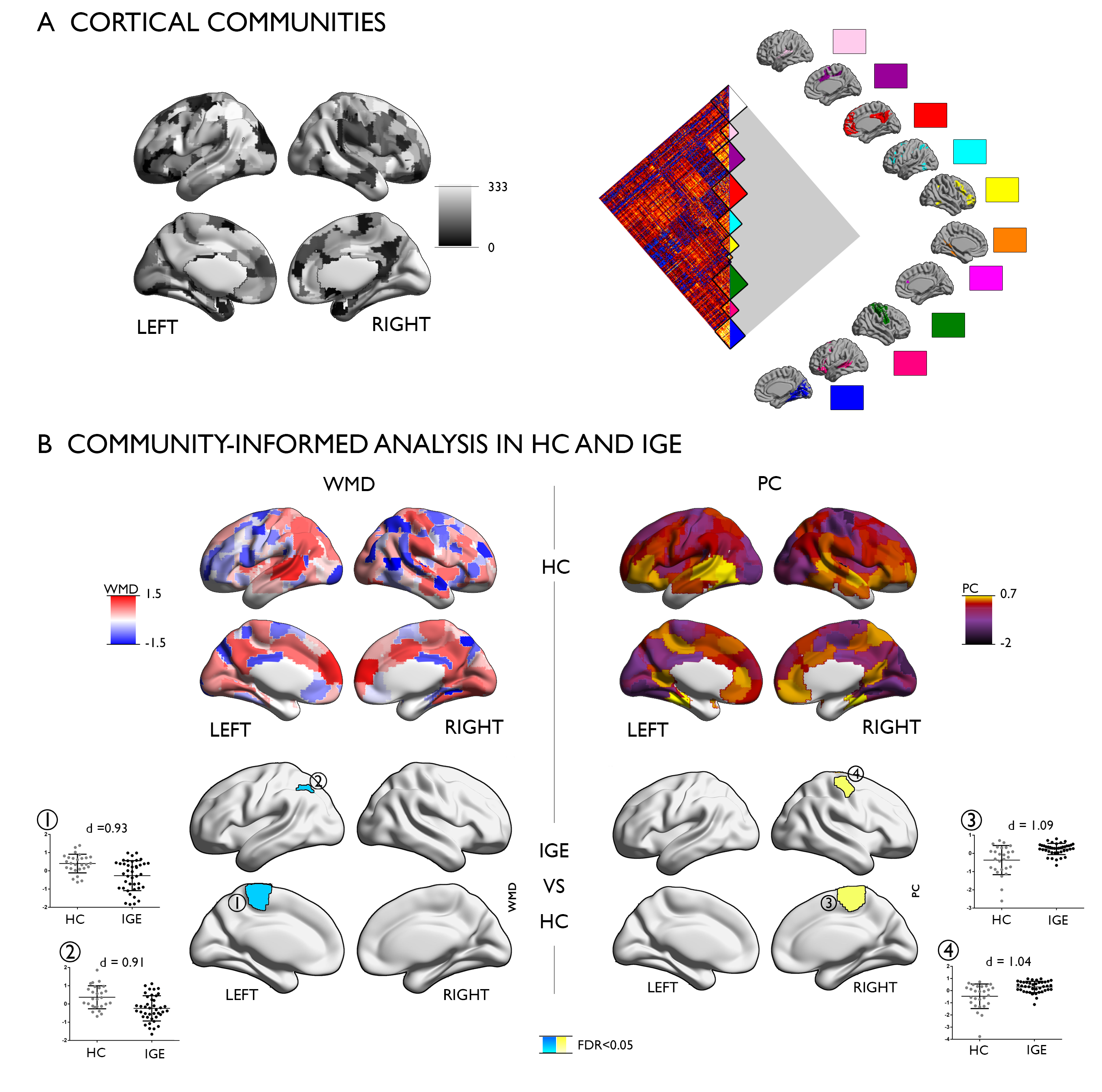
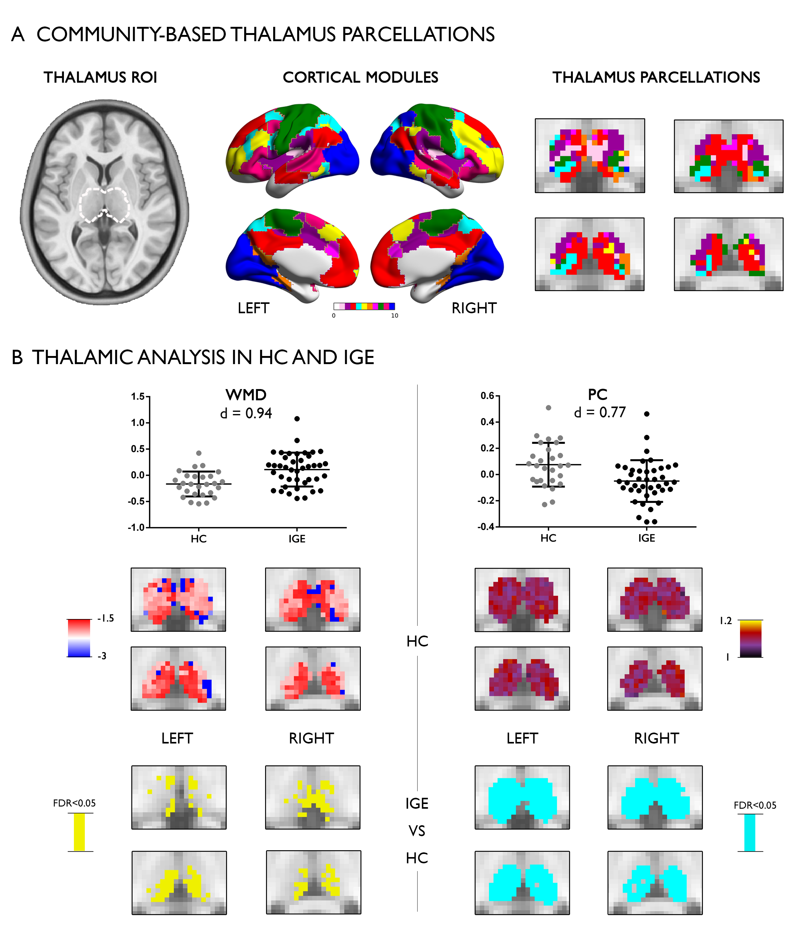
We studied 42 IGE-GTCS prospective patients and 27 healthy controls (HC) using resting-state BOLD fMRI (TR = 2000ms, TE = 30ms, flip angle = 90°, voxel size = 3.0×3.0×4.0 mm3, 35 slices, 230 volumes) and T1-weighted structural MRI (TR = 9.8ms, TE = 4.6ms, TI = 900ms, flip angle = 8°, voxel size = 1.0×1.0×1.0 mm3, 192 slices) on a Philipps 3T MRI system. After 1 year of follow-up, 14 patients were classified as seizure-free and 28 were drug-resistant. We subdivided the neocortex into 333 parcels and computed cortico-cortical and thalamo- cortical connectomes. We grouped cortical parcels into large-scale communities and assigned thalamic voxels to cortical communities using a winner-take-all scheme. For each cortical parcel and thalamic voxel, we calculated the participation coefficient, PC, a measure of between-network connectivity, and the within-module degree, WMD, to index within-network connectivity. PC and WMD measures were z-normalized relative to their community, and we compared PC and WMD measures between IGE-GTCS and controls, and between seizure-free and drug-resistant patients. FDR adjustment corrected for multiple comparisons. Connectome analysis was used to assess connectivity within and between large-scale network communities in both cortical and thalamic subregions. In addition to comparing patients to controls, we examined associations to prospective seizure control.Results
In the cortico-cortical connectome (Figure 1), HC showed high WMD and PC in bilateral medial prefrontal, medial parietal, and insular regions, together with left temporo-parietal regions. WMD was furthermore elevated in bilateral medial occipital regions, while PC was high in lateral prefrontal cortices. Comparing IGE to HC at the level of cortico-cortical connectivity revealed reductions in WMD in patients, while PC was increased. Specifically, patients showed lower WMD in the left paracentral lobule and angular gyrus (p<0.05, FDR-corrected). In contrast, they showed increased PC in right fronto-central (p<0.05, FDR-corrected).Assessing thalamo-cortical WMD and PC (Figure 2), we observed high WMD and PC in anterior, medial, posterior, and dorsal thalamic divisions in HC. Comparing thalamo-cortical WMD and PC between IGE and HC, patients demonstrated marked increases of WMD (p<0.05, FDR-corrected) and a widespread reduction in PC (p<0.05, FDR-corrected). Comparing mean cortical thickness between IGE and HC, we did not find significant differences between both groups. Similarly, vertex-wise cortical thickness comparisons did not result in any findings that survived correction for multiple comparisons. On the other hand, IGE showed bilateral thalamic atrophy (p<0.025). Importantly, functional network alterations in IGE compared to HC were robust when additionally controlling for mean cortical thickness in the statistical models, including WMD decreases and PC increases in cortical regions in patients. Similarly, thalamic WMD increases and PC decreases in IGE compared to HC were robust when controlling for thalamic volume. We stratified patients based on their drug-response patters after 1 year of follow-up into seizure-free and drug-resistant, controlling for age, sex, and duration of epilepsy. Comparing both IGE subgroups, we observed more marked thalamo-cortical imbalances in patients who were diagnosed as drug-resistant compared to seizure free patients. Specifically, we observed lower WMD in left paracentral lobule and higher PC in the left angular gyrus (p<0.05) as well as increased WMD in the thalamus in drug-resistant patients (p<0.05).
Conclusions
Our findings suggest a pathoconnectomic substrate of IGE centered on diverging changes in cortical and thalamic connectivity. More restricted thalamic connectivity could reflect the tendency to engage in recursive thalamo-cortical loops, which may contribute to network hyper-excitability. Conversely, increased connectional diversity of cortico-cortical networks may relay abnormal activity to an extended bilateral cortical territory. Network imbalances related to future drug-response, suggesting potential for prognostic applications.
Acknowledgements
We acknowledge funding from CIHR, FRQS, NSFC.References
No reference found.Figures