3119
Comparative analysis of Mean Apparent Propagator (MAP)-MRI and traditional DTI for the diagnosis of hippocampal sclerosis in temporal lobe epilepsy1The First Affiliated Hospital of Zhengzhou University, Zhengzhou, China, 2MR Scientific Marketing, Siemens Healthcare, Shanghai, China, Shanghai, China
Synopsis
This study aimed to preliminarily investigate the utility of MAP-MRI parameters in hippocampal sclerosis and compare them with traditional DTI parameters to explore the changes in hippocampal microstructure and understand its pathophysiological mechanisms. The swelling of the hippocampal nerve fiber axon presynaptic terminal can be reflected by MAP-MRI, which greatly assists in the clinical diagnosis of early hippocampal sclerosis. Compared with DTI parameters, DSI parameters showed higher diagnostic efficacy particularly for RTPP and QIV. This study extracted more objective and reproducible parameters of fixed hippocampal sclerosis. More samples are needed for future research.
Introduction
Abnormalities of medial temporal lobe structures (hippocampus, amygdala, inner olfactory cortex, etc.) can cause medial temporal lobe epilepsy. Hippocampal sclerosis (HS) may be one of the most important causes of epilepsy. Partial anterior temporal lobe, hippocampus, and hippocampal resection can control more than 70% of refractory temporal lobe seizures. Therefore, accurate positioning of preoperative lesions is a key factor for successful surgical outcomes.
The diffusion spectrum imaging (DSI) technique can provide information on the dispersion of water molecules, which might reflect microstructural changes in brain tissue. Recently, a DSI-based model called mean apparent propagation (MAP)-MRI was introduced and could derive quantitative parameters from the probability density function (PDF) of water motion1. Application of the MAP-MRI method in hippocampal sclerosis has not yet been reported in the literature. This study aimed to investigate the value of MAP-MRI for the diagnosis of hippocampal sclerosis and compare it with diffusion tensor imaging (DTI) to screen out more favorable parameters to improve its clinical diagnosis.
Method
Thirteen patients (mean age: 30.7 ± 14.1 years; median age: 28 years) with unilateral hippocampal sclerosis (left: 5 cases; right: 10 cases) confirmed by surgical pathology were enrolled in the study. All the patients underwent MRI acquisition on a 3T scanner (MAGNETOM Prisma, Siemens Healthcare, Erlangen, Germany), including the oblique T2 FLAIR sequence perpendicular to the hippocampus, DTI, and DSI. The DSI images were acquired using a q-space sampling scheme, with the q-space weighting 4 and half coverage. Two different b-values (0 and 3000 s/mm2) were included in the acquisition. The other sequence parameters were as follows: TR/TE = 3800/72 ms, FOV = 220 × 220 mm2, GRAPPA = 2, slice acceleration factor = 2, slice thickness = 2.2 mm, voxel size = 2.0 × 2.0 × 2.2 mm3, slices = 60, and scan time=4.45min. The DTI and MAP-MRI parameters were estimated from q-space diffusion data using software developed in-house, which is based on an open-resource tool DIPY (Diffusion Imaging In Python, http://nipy.org/dipy). The DTI parameters included axial diffusivity (AD), the fractional anisotropy (FA), primarily the mean diffusivity (MD), and radial diffusivity (RD). The DSI parameters included the mean square displacement (MSD), non-Gaussian parallel (NG∥), non-Gaussian perpendicular (NG⊥),q-space inverse variance (QIV), the return to the origin probability (RTOP), the return to the axis probability (RTAP), and the return to the plane probability (RTPP).
The region of interest (ROI) was drawed in each side of hippocampus of the patients separately, and the hippocampal sclerosis side was compared with the normal side. The optimal threshold value was determined by plotting the receiver's operating characteristic (ROC) curve, and the discriminant efficacy of each parameter was calculated. The ROC curves were compared in pairs by the Delong method. For all the statistical analyses, P < 0.05 was considered to be significant.
Results
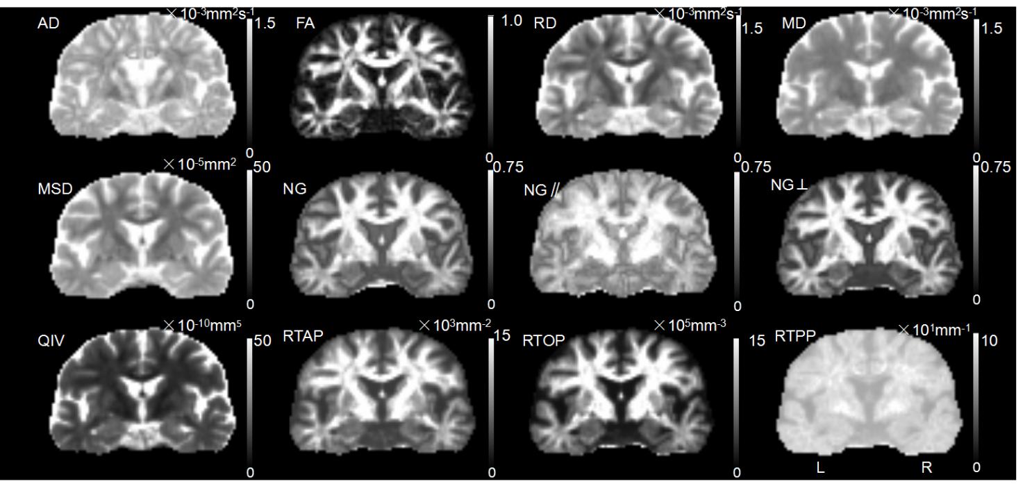
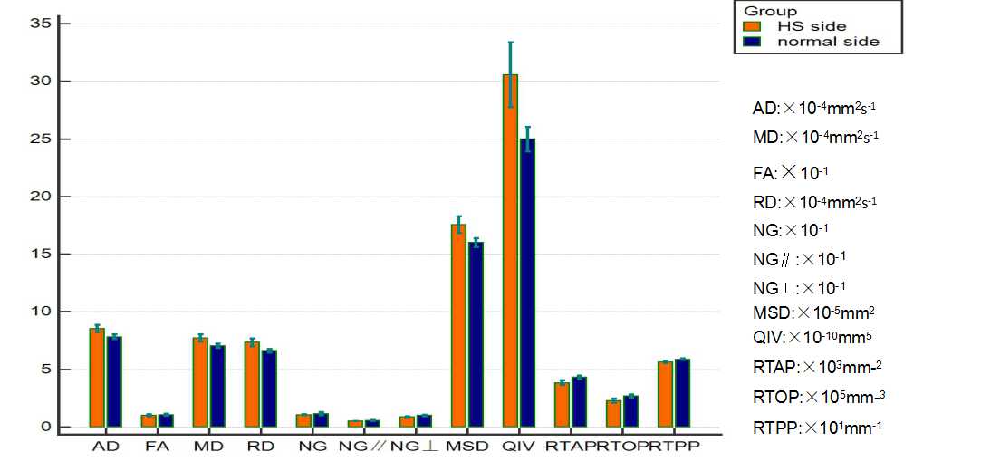
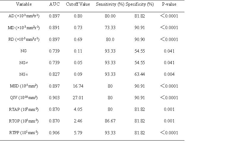
Figure 1 shows a patient’s map of the DTI and MAP-MRI parameters. The mean values of AD, MD, RD, MSD, and QIV in ROI for the hippocampal sclerosis side were higher than those of the normal side (P < 0.05), and the mean values of NG, NG∥, NG⊥, RTAP, RTOP, and RTPP were lower than those of the normal side (P < 0.05), as shown in Table 1 and Figure 2. The mean values of FA showed no significant difference between the two groups (P > 0.05).The diagnostic performance of the statistically significant parameters is shown in Table 1. RTPP had a dominantly high AUC (0.906) in the diagnosis of hippocampal stenosis . NG, NG∥, NG⊥, and RTPP showed higher sensitivity (86.67% – 93.33% vs. 73.33 - 80.00%, p < 0.05) but lower specificity (54.55% – 81.82% vs. 90.91 - 90.90%, p < 0.05) than MD and RD for the diagnosis of hippocampal sclerosis.Discussion
The typical manifestations of hippocampal sclerosis are glial cell proliferation and neuronal loss that is more pronounced in vulnerable epileptic areas. The quantitative parameters from the MAP-MRI model showed signal changes in the microstructure of the hippocampus, suggesting associated pathological changes, which may reflect the swelling of hippocampal mossy fibers and could provide imaging evidence for the early diagnosis of HS2,3.There was no statistical significance in FA for the DTI parameters. However, some parameters of DTI, such as MD and AD, have high specificity, suggesting that two methods can be combined to help diagnose early hippocampal sclerosis in clinical work. Considering the possibility of bias caused by a smaller sample size, more samples need to be studied.Acknowledgements
No acknowledgement found.References
1.Ozarslan E, Koay C G, Shepherd T M, et al. Mean apparent propagator (MAP) MRI: a novel diffusion imaging method for mapping tissue microstructure. Neuroimage.2013;78:16-32.
2.Boscolo G I, Brusini L, Obertino S, et al. On the Viability of Diffusion MRI-Based Microstructural Biomarkers in Ischemic Stroke. Front Neurosci. 2018;12:92.
3.Avram A V, Sarlls J E, Barnett A S, et al. Clinical feasibility of using mean apparent propagator (MAP) MRI to characterize brain tissue microstructure. Neuroimage, 2016;127:422-434.
Figures