3108
Ex-vivo diffusion MRI of human hemispheres: the effect of tissue fixation and the relation to in-vivo diffusion MRI1Rush Alzheimer's Disease Center, Rush University Medical Center, Chicago, IL, United States, 2Department of Biomedical Engineering, Illinois Institute of Technology, Chicago, IL, United States, 3Department of Neurological Sciences, Rush University Medical Center, Chicago, IL, United States, 4Department of Diagnostic Radiology, Rush University Medical Center, Chicago, IL, United States
Synopsis
Until the relationship between in-vivo and ex-vivo diffusion measures has been established, the clinical relevance of findings using ex-vivo diffusion MRI is unclear and precludes potential studies. Therefore, this study investigated the effects of tissue fixation on basic diffusion measures, and established the relationship of diffusion measures recorded in-vivo and ex-vivo. Basic diffusion measures of postmortem hemispheres were observed over 5 weeks. The relationship between in-vivo and ex-vivo diffusion measures was studied using linear mixed regression. Appreciable changes to diffusion measures were seen early on in fixation, and ex-vivo measurements of FA and RD were linked to their in-vivo measurements.
Introduction
Combining ex-vivo MRI with pathology can provide valuable information in understanding age-related diseases; This has been observed for R2 relaxation and volumetry. Ex-vivo diffusion MRI may be linked to age-related neuropathology; However, until the relationship between in-vivo and ex-vivo diffusion measures has been established, the clinical relevance of findings using ex-vivo diffusion MRI is unclear and precludes potential studies. Therefore, the objectives of this work were to investigate the effects of tissue fixation on basic diffusion measures, and to establish the relationship of diffusion measures recorded in-vivo and ex-vivo.Methods
Participants:
Nine participants were recruited from the Rush Memory and Aging Project, and the Religious Orders Study, two longitudinal, clinical-pathologic cohort studies of aging. Participants signed an anatomical gift act, and upon death, the participants’ postmortem hemisphere was extracted and submerged in 4% formaldehyde for imaging.
Data collection:
Two datasets were used. The effect of tissue fixation on diffusion measures was evaluated with Dataset 1, and the relationship between in-vivo and ex-vivo diffusion measures was established with Dataset 2.
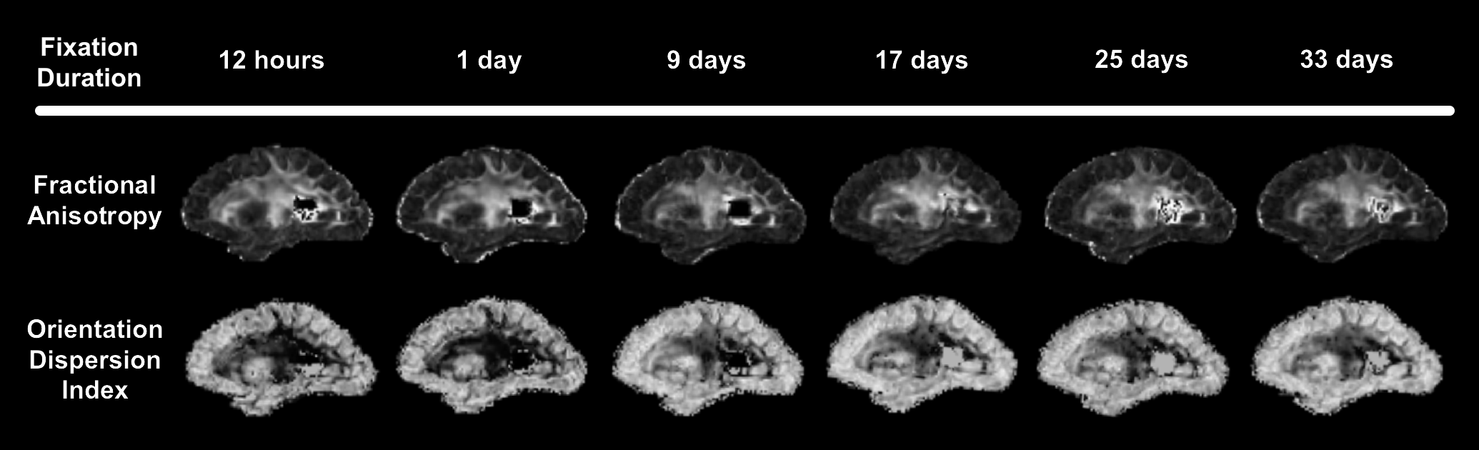
-Dataset 1: The postmortem hemispheres of five participants were imaged at multiple time points: immediately after death, 1 day after death, and on a weekly basis for 5 weeks. Ex-vivo diffusion data were collected on a 3T Philips scanner with multi-shell acquisition (b=2000 to 6000, increments of 1000; and 25 directions).
-Dataset 2: Four participants were imaged both in-vivo and ex-vivo. In-vivo DTI data were collected biennially until death on a 3T Siemens scanner using a single-shell acquisition (b=1000 and 45 directions). In-vivo data most proximal to death was used. Ex-vivo diffusion data were collected early after death (<24 hrs.) on a 3T Philips scanner with a single-shell acquisition (b=3000 and 125 directions).
Effect of tissue fixation on diffusion measures (Dataset 1):
At each time point, fractional anisotropy (FA), axial diffusivity (AD), and radial diffusivity (RD), were calculated with the free water elimination model in DIPY. Additionally, intra-cellular volume fraction (ICVF) and orientation dispersion index (ODI) were obtained with NODDI.
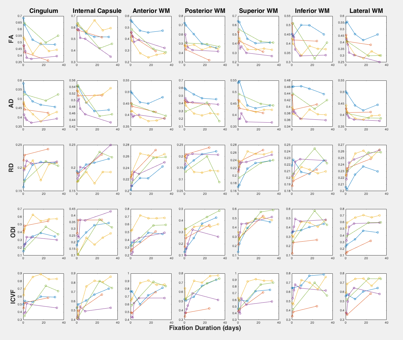
Using DTITK, a template was generated for each participant, and then the participants’ templates were used to generate a group template. The cingulate, internal capsule, and five white matter areas located in the anterior, posterior, superior, inferior, and lateral sections were manually outlined on the group template. The mean values for the above diffusion measures were recorded for all regions and time points.
For each DTI measure, the measurements were regressed with fixation duration using a generalized linear mixed model with random effects of participant and region. Regional values of AD and RD were normalized by trace. Significance was achieved at p<0.05.
Relationship between in-vivo and ex-vivo DTI measures (Dataset 2):
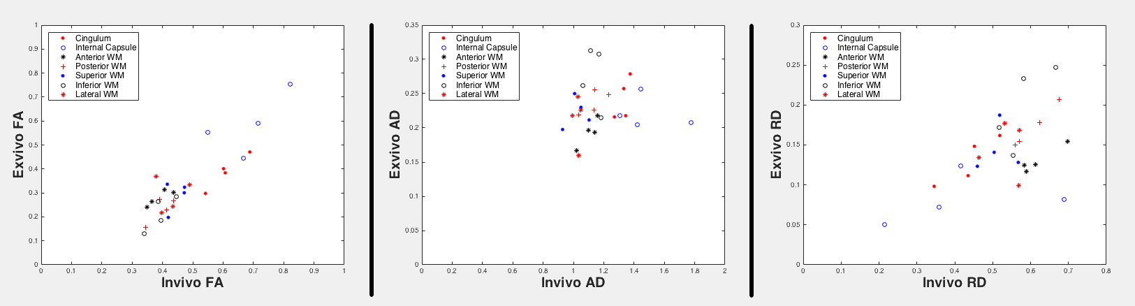
Since Dataset 2 relies on single-shell data, FA, AD, and RD were calculated with the tensor model, and ODI and ICVF were not calculated. The postmortem hemisphere of each participant was registered to the corresponding antemortem hemisphere.
A group template was generated using the whole brain in-vivo data, and the same regions above were manually outlined on the group template. The mean values for the diffusion measures were recorded for both in-vivo and ex-vivo in all regions.
Ex-vivo measurements were regressed with their corresponding in-vivo measurements using linear mixed modeling with random effects of participant and region. Significance was achieved at p<0.05.
Results
Generalized linear mixed modeling showed that FA and normalized AD decreased as the tissue was fixed (pFA=3.2e-16, pnAD=3.3e-6), while on the other hand, RD, ODI and ICVF increased with fixation (pnRD=1.3e-10, pODI=3.0e-14, pICVF=4.8e-18). Most participants had appreciable changes in their diffusion measures within the first 10 days (Fig. 1, Fig. 2). In the analysis of the in-vivo and ex-vivo relationship, in-vivo measurements of FA and RD have significant associations with their corresponding ex-vivo measurements (pFAvivo=2.7e-8, pRDvivo=2.8e-2) (Fig. 3).Discussion and Conclusions
All diffusion measures in this study (FA, normalized AD, normalized RD, ICVF, and ODI) were significantly associated with fixation duration, and noticeable changes to these diffusion measures were observed early on in fixation (within the first 10 days). This finding suggests that ex-vivo diffusion MRI studies should utilize MRI data collected soon after death to minimize the effects of tissue fixation on the results. Within the same participants, ex-vivo measurements of FA and RD were related to their corresponding in-vivo measurements, providing clinical relevance to prospective ex-vivo MRI and pathology studies.Acknowledgements
NINDS UH2NS100599References
No reference found.Figures