3099
Magnetic resonance spectroscopic imaging of hyperpolarized [1-13C]pyruvate in glioblastoma: a promising tool for investigating tumor metabolism and heterogeneity.1Department of Radiology, University of Cambridge, Cambridge, United Kingdom, 2Cancer Research UK Cambridge Institute, University of Cambridge, Cambridge, United Kingdom, 3GE Global Research, Munich, Germany, 4Department of Pathology, Cambridge University Hospitals NHS Foundation Trust, Cambridge, United Kingdom, 5Cambridge Cancer Centre, Cambridge University Hospitals NHS Foundation Trust, Cambridge, United Kingdom, 6Department of Radiology, Cambridge University Hospitals NHS Foundation Trust, Cambridge, United Kingdom, 7Department of Medicine, University of Cambridge, Cambridge, United Kingdom, 8Department of Oncology, Cambridge University Hospitals NHS Foundation Trust, Cambridge, United Kingdom, 9Department of Clinical Neurosciences, University of Cambridge, Cambridge, United Kingdom
Synopsis
Glioblastomas (GBM) are characterized by diffuse infiltration, a high level of intratumoral and intertumoral heterogeneity and a very poor prognosis. Characterising tumor heterogeneity in vivo may improve diagnosis, therapy planning and treatment assessment. Dissolution dynamic nuclear polarization (DNP) is a novel technique that allows dynamic and non-invasive assessment of the metabolism of hyperpolarized (HP) 13C-labelled molecules in vivo, such as the preferential exchange of [1-13C]pyruvate to [1-13C]lactate within tumors (Warburg effect). In this study we explore metabolic reprogramming within glioblastoma (GBM) and its microenvironment using HP [1-13C]pyruvate to demonstrate the heterogeneity of pyruvate’s metabolic fate.
Introduction
Glioblastomas (GBM) are extremely heterogeneous, both morphologically and biologically, which contributes to a very poor prognosis. Metabolic reprogramming is one of the major driving forces in determining this behavior, therefore the characterization of metabolic intratumoral, peritumoral and intertumoral heterogeneity in vivo could help to develop novel therapeutic strategies targeting cancer-specific metabolic pathways1. 13C magnetic resonance imaging using dynamic nuclear polarization (DNP) is a novel technique that allows for non-invasive assessment of the metabolism of hyperpolarized (HP) 13C-labelled molecules in vivo, such as the preferential conversion of [1-13C]pyruvate to [1-13C]lactate in tumors (Warburg effect). The purpose of this study was to explore metabolic reprogramming in glioblastoma and the surrounding microenvironment using HP [1-13C]pyruvate.Methods
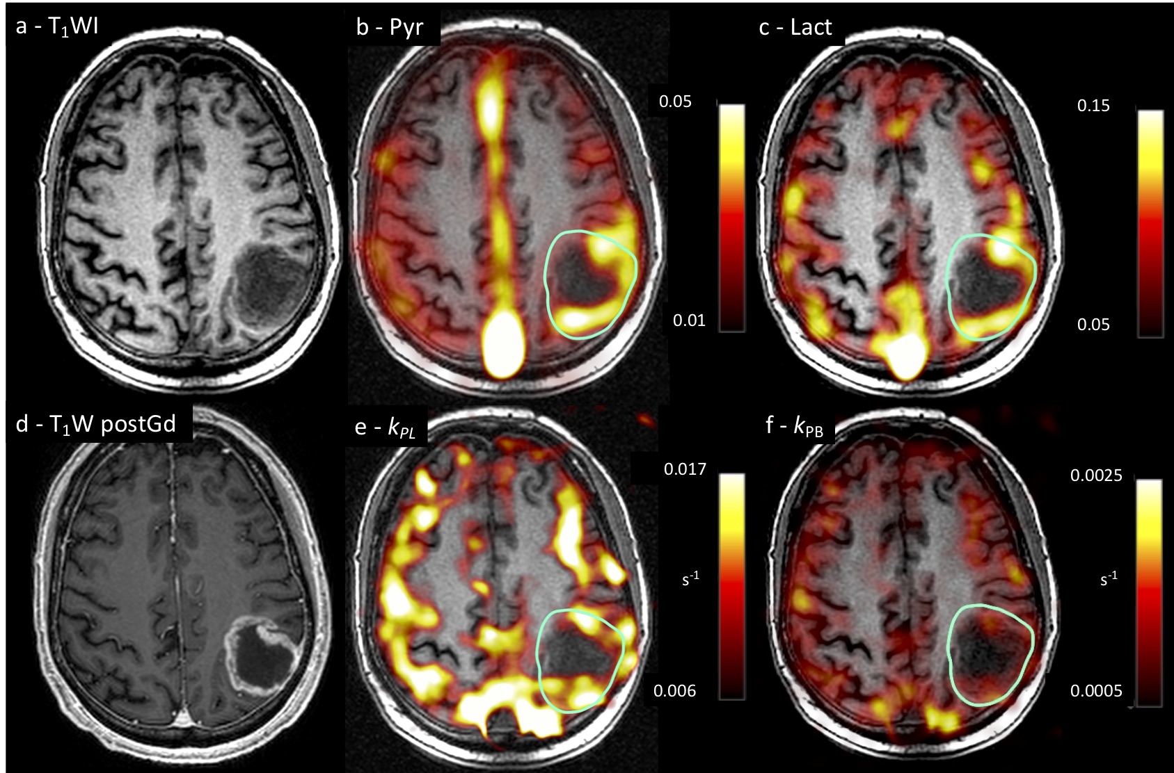
Three treatment-naïve patients (1 male, 2 female, 58.3 ±12.9 years) with GBM were imaged on a 3T scanner (MR750, GE Healthcare, Waukesha, WI, USA) using a dual-tuned 13C/1H quadrature transmit/receive head coil (Rapid Biomedical, Rimpar, Germany). [1-13C]pyruvate was hyperpolarized using a clinical hyperpolarizer (Research Circle Technology, Albany NY). 13C MRSI acquisition was performed using a dynamic IDEAL spiral sequence2 (FOV=24 cm, TR=500, Flip angle=15°, acquisition time=60s, temporal resolution=4s, acquisition matrix=40x40, reconstruction matrix=128x128, slice thickness=3cm). Apparent kinetic rate constant maps for the conversion of pyruvate to lactate (kPL) and pyruvate to bicarbonate (kPB) were derived using a two-site model3. A reference 3D T1-weighted Fast SPoiled GRadient echo (T1W FSPGR; FOV=24cm, TR=8.6, TE=3.2, acquisition matrix=256x224, reconstruction matrix=256x256; slice thickness=2mm) was acquired with the dual-tuned 13C/1H quadrature head coil. After the [1-13C]pyruvate acquisition, a T1W FSPGR (FOV=24cm, TR=8.2, TE=3.2, acquisition matrix=256x256, reconstruction matrix=256x256; slice thickness=1mm) was acquired using a 1H 12-channel head coil following the injection of a gadolinium-based contrast agent (Gadobutrol 1.0 mmol/mL; Schering). Regions of interest (ROIs) were obtained for whole tumor, peritumoral region, non-enhancing and contrast-enhancing regions on the unenhanced T1W FSPGR using the post gadolinium injection sequence as guidance. For comparison, the normal appearing brain parenchyma (NABP) was assessed in each hemisphere, each lobe and at the level of the basal ganglia. To assess the spatial heterogeneity within the lesion, regional analysis was performed selecting multiple ROIs in the medial, lateral, anterior and posterior aspects of the lesion as well as from the middle. An unpaired t-test was used to compare the kinetic values derived from the GBM and the NABP; significance was assessed at p<0.05.Results
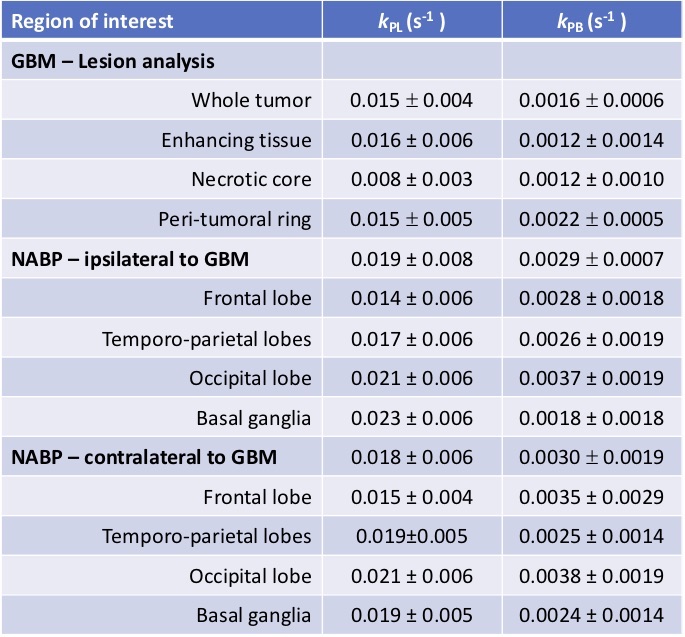
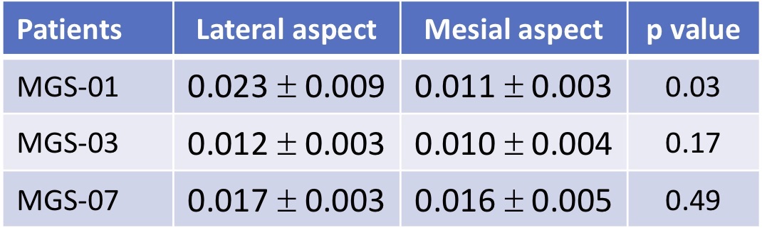
Dynamic signal from hyperpolarized pyruvate, lactate, and bicarbonate were observed in the brain following intravenous injection of [1-13C]pyruvate. A representative case is shown in figure 1. Kinetic analysis (table 1) demonstrated a significantly lower kPL in whole tumor compared to the contralateral NABP (p = 0.03) but not compared to the ipsilateral NABP (p = 0.05). Similarly, the kPB in glioblastoma was significantly lower compared to both the contralateral NABP (p < 0.0001) and the ipsilateral NABP (p < 0.0001). In the peritumoral region, kPL was not significantly different from the contralateral NABP (p = 0.22), however the peritumoral kPB was lower than in the contralateral NABP (p = 0.017). The regional analysis demonstrated a trend of higher kPL in the lateral aspect of the region (table 2); however, only in one patient was the difference statistically significant (p = 0.03). No adverse events were observed in patients following injection of [1-13C]pyruvate.
Discussion
This study demonstrates the conversion of hyperpolarized pyruvate to both lactate and bicarbonate in the human brain and in glioblastoma. GBMs demonstrate marked intratumoral and intertumoral variation and this heterogeneity is also seen on a metabolic level in this study. The exchange of hyperpolarized 13C label between pyruvate and lactate (kPL) was lower in GBM compared to the NABP. The unidirectional formation of bicarbonate (KPB) was also consistently reduced within GBM compared to NABP. A decrease in both these parameters suggests a reduction in viable cell density within the tumor and metabolic remodelling. Moreover, we were able to detect intralesional heterogeneity in the labelling exchange highlighting the presence of different metabolic habitats that could represent targets for image-guided biopsies.
These results could deepen our understanding of metabolic reprogramming in GBM and the surrounding brain in vivo, which may enable better stratification, tumor staging, and better monitoring of therapeutic response. Future work will include a larger cohort to correlate hyperpolarized 13C metabolic imaging with histopathology derived at surgery.
Conclusion
HP 13C MRSI can probe in vivo metabolic activity of GBM and investigate both intratumoral and intertumoral heterogeneity.Acknowledgements
This work has been funded by Cancer Research UK (CRUK; C19212/A16628) and the CRUK & Engineering and Physical Sciences Research Council (EPSRC) Cancer Imaging Centre in Cambridge and Manchester (C197/A16465). Additional support has been provided by the CRUK Cambridge Centre, the National Institute of Health Research (NIHR) Cambridge Biomedical Research Centre and Addenbrooke’s Charitable Trust. The views expressed are those of the author(s) and not necessarily those of the NHS, the NIHR or the Department of Health and Social Care.References
1. Corbin Z, Spielman D, Recht L. A Metabolic Therapy for Malignant Glioma Requires a Clinical Measure. Curr. Oncol. Rep. 2017;19(12).
2. Wiesinger F, Weidl E, Menzel MI, et al. IDEAL spiral CSI for dynamic metabolic MR imaging of hyperpolarized [1-13C]pyruvate. Magn. Reson. Med. 2012;68(1):8–16.
3. Khegai O, Schulte RF, Janich MA, et al. Apparent rate constant mapping using hyperpolarized [1-13C]pyruvate. NMR Biomed. 2014;27(10):1256–1265.
Figures