3096
Fast Field-Cycling MRI identifies ischaemic stroke at ultra-low magnetic field strength1Aberdeen Biomedical Imaging Centre, University of Aberdeen, Aberdeen, United Kingdom, 2Acute Stroke Unit, Aberdeen Royal Infirmary, Aberdeen, United Kingdom
Synopsis
In this work we present the first patient images from our home built Fast Field-Cycling MRI (FFC-MRI) scanner. By varying the external magnetic field during the imaging process, FFC-MRI allows us to probe the variation of T1 with magnetic field, known as “T1 dispersion”. This T1 dispersion has potential value as a diagnostic biomarker in a range of conditions. Here we present images demonstrating that endogenous T1 contrast at 20 mT and below can be used to identify ischaemic stroke.
Introduction
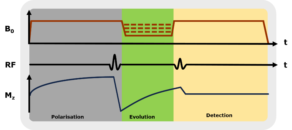
Fast Field-Cycling MRI1 (FFC-MRI) is a novel MRI technique in which the external magnetic field is switched rapidly between levels during the imaging experiment. In this way, FFC-MRI gains access to information which is invisible to conventional MRI scanners, especially the variation of T1 with magnetic field, known as “T1 dispersion”. A typical field-cycling experiment is comprised of three periods (Fig. 1): polarisation, evolution and detection. During polarisation, B0 is set at the maximum value for a period of time, typically 1 - 5 times T1, in order to build up a sufficiently large value of Mz. Next the magnetic field is switched to a magnetic field of interest – the evolution field – for a time period on the order of T1, at which the system will undergo spin-lattice relaxation. Finally the system is returned to the detection field, at which the signal is read out using conventional MRI. By repeating the experiment for various values of evolution fields, images displaying T1 at different field strengths can be obtained, contributing to a T1-dispersion curve. Polarising using a relatively large value of B0, (e.g. 0.2 T) allows evolution fields of 1 mT and below to be probed without significant loss of SNR. The T1 values corresponding to these ultra-low magnetic fields are associated with slow molecular motion with long correlation times, which may have diagnostic value in a wide range of pathologies. In this work we aimed to assess whether we could identify recent cerebral infarcts at ultra-low field strength, when compared with conventional imaging.
Methods
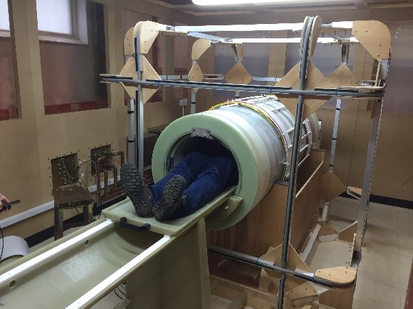
After informed consent, a group of patients (n = 22) with ischemic stroke were scanned using FFC-MRI within 24-96 h of presentation. Initial diagnosis was performed using CT and/or 3T MRI. The FFC-MRI scans were performed using a home-built field-cycling scanner (Fig 2) comprised of a resistive magnet with a maximum field strength of 0.2 T. Sets of images from five different evolution fields ranging from 0.2 T to 0.2 mT were obtained using a spin-echo readout. The FFC-MRI imaging parameters were: Matrix size 128 x 128, FOV = 280 mm, THK = 10 mm, TE = 24 ms, NEX = 1. Total examination duration, including setup time, was approximately 45 minutes.Results
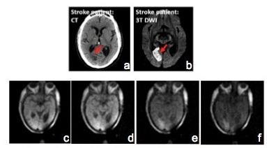
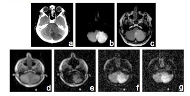
In patients with sub-acute ischaemic stroke, T1-weighted FFC-MRI images exhibited hyper-intense regions, with contrast increasing markedly as the evolution magnetic strength field decreased, with maximum contrast at the lowest field used (0.2 mT). The infarct region measured by FFC-MRI correlated well with the abnormality in CT and/or DWI images (Examples in Fig 3,4).Discussion
This is the first-ever clinical application of this new modality, proving that FFC-MRI can generate diagnostic-quality images of ischaemic stroke at ultra-low magnetic fields (e.g. 0.2 mT), with significantly enhanced endogenous T1-contrast compared to conventional MRI. These findings have implications for future development of a new and safe imaging modality not only for stroke but also for many other clinical conditions.Acknowledgements
This project has received funding from the European Union’s Horizon 2020 research and innovation programme under grant agreement No 668119 (project “IDentIFY”).References
Lurie, D. J. et al. Fast field-cycling magnetic resonance imaging. Comptes Rendus Phys. 11, 136–148 (2010).Figures