3093
Enhancing Fluorine-19 Magnetic Resonance Drug Imaging: Chemical Variations in the Fluorine Side-Groups of the Immunomodulatory Drug Teriflunomide1Berlin Ultrahigh Field Facility, Max Delbrück Center for Molecular Medicine, Berlin, Germany, 2Departments of Chemical Biology and Structural Biology, Leibniz-Institut fϋr Molekulare Pharmakologie (FMP), Berlin, Germany, 3Screening Unit, Leibniz-Institut für Molekulare Pharmakologie (FMP), Berlin, Germany, 4Experimental and Clinical Research Center, a joint cooperation between the Charité Medical Faculty and the Max Delbrück Center for Molecular Medicine, Berlin, Germany
Synopsis
Fluorine-19 (19F)-MR is of high relevance for the study of fluorinated drugs in vivo. Due to low drug concentrations and low numbers of fluorine atoms per molecule, the signal to be detected is very low. To address this drawback this work enhances 19F MRI of the antiinflammatory drug teriflunomide. For this purpose, derivatives of the trifluorinated drug were synthesized including modifications of the number and position of fluorine atoms in the 19F side-chains. We studied the 19F NMR characteristics and compared the SNR efficiencies of these compounds. The inhibitory activity was studied and correlated with the detectability of the compounds. By this, we can select drugs which provide a better signal than the original teriflunomide and which show an equal or even better biological activity.
Introduction
Fluorine-19 (19F) magnetic resonance (MR) techniques are commonly used for studying the biodistribution of fluorinated drugs1. The achievable signal-to-noise-ratio (SNR) is limited, due to the low availability of 19F drugs in vivo as well as the low number of 19F atoms per molecule, when compared to 19F nanoparticles used for cell tracking during inflammation2. Teriflunomide (TF) is a trifluorinated anti-inflammatory drug to treat Multiple Sclerosis inhibiting the mitochondrial enzyme dihydroorotate dehydrogenase (DHODH)3. To enhance the 19F MR signal that can be detected in vivo, novel derivatives of TF were synthesized in this work. For this purpose, the 19F-sidechains of TF were modified with respect to the position and number of 19F atoms. We studied the 19F NMR properties (spectrum, relaxation times) and compared the SNR efficiencies of TF and four of its derivatives with their DHODH inhibitory activity using a colorimetric enzyme activity assay. The aim was to improve the MR detectability of DHODH inhibitors while preserving their inhibitory activity.Methods
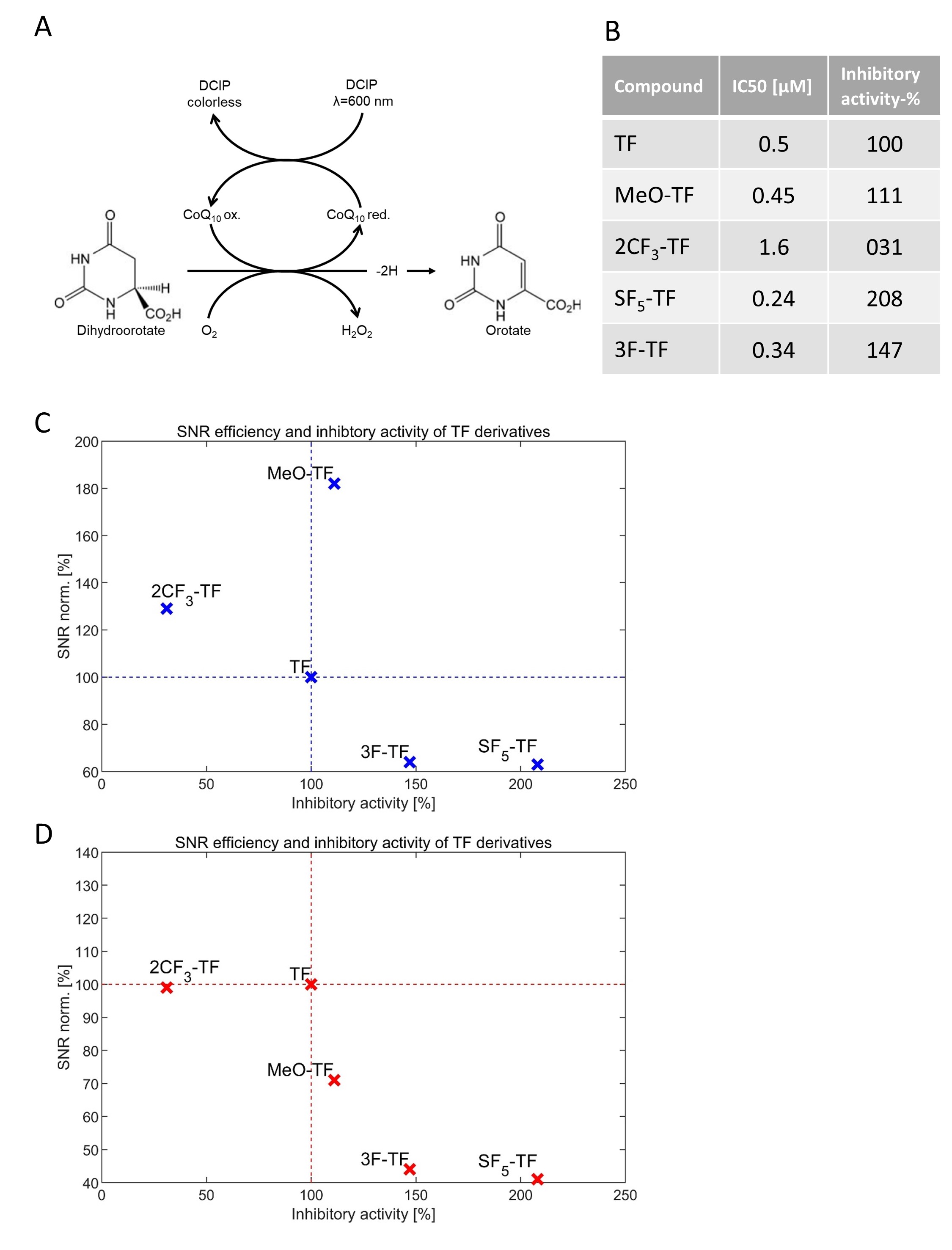
All derivatives were prepared in 2ml syringes. The concentration of the compounds was adjusted to 6.56E+16 19F atoms/ml. All MR experiments were performed on a 9.4T MR scanner (Bruker Biospec, Ettlingen, Germany) using a dual-tunable 19F/1H mouse head RF coil2. MR measurements were performed at room temperature (RT) and at 37°C. Global single pulse spectroscopy (TR=1000ms, TA=8s) was used to detect the 19F signal and to make frequency adjustments. T1 mapping was performed using RARE (TE=4.6ms, ETL=4, FOV=16mmx16mm, matrix size=64x64, with 9 variable repetitions times (TR=25ms-8000ms). T2 mapping was performed using a multi-slice multi-echo sequence (TR=2000ms, FOV=16mmx16mm, matrix size=64x64) with 25 different TEs (TE=40-1000ms in steps of 40ms for long T2, TE=8-200ms in steps of 8ms for short T2). RARE (FOV=16mmx16mm, matrix size=64x64) was optimized (TR, TE and ETL versus relaxation times4) to achieve highest possible SNR for the assessment of the SNR efficiency (SNR/sqrt(time)) of the compounds. The inhibitory activity (drug concentration range: 50µM to 5nM) was examined by monitoring the reduction of 2,6-dichloroindophenol (DCIP) by the loss of absorbance at 620nm5, which is associated with oxidation of L-dihydroorotate (L-DHO) catalyzed by the DHODH enzyme6. The reaction was performed in Tris-HCl buffer, containing coenzyme Q10, Triton X-100, KCl, DCIP, and DHODH.Results
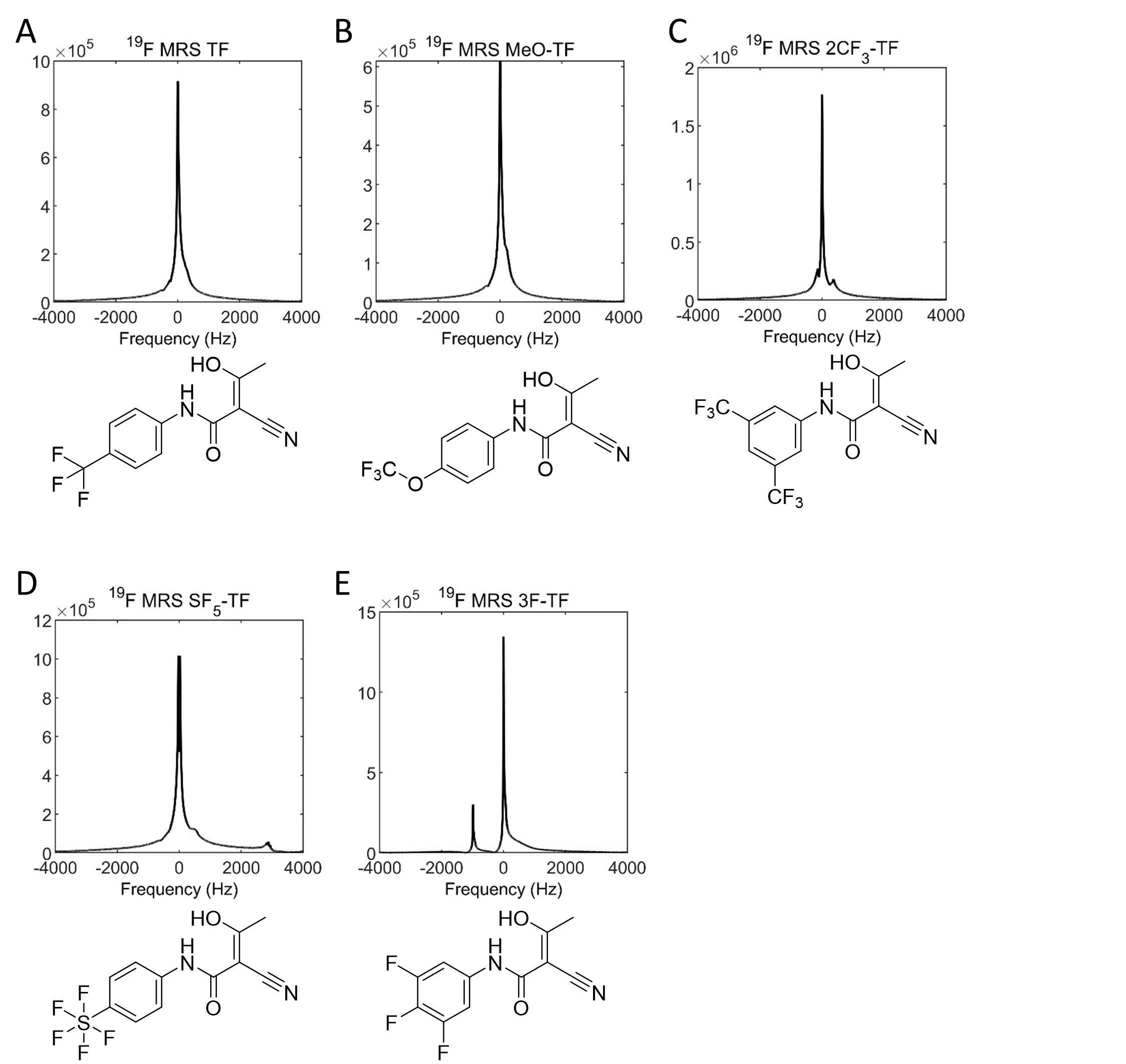
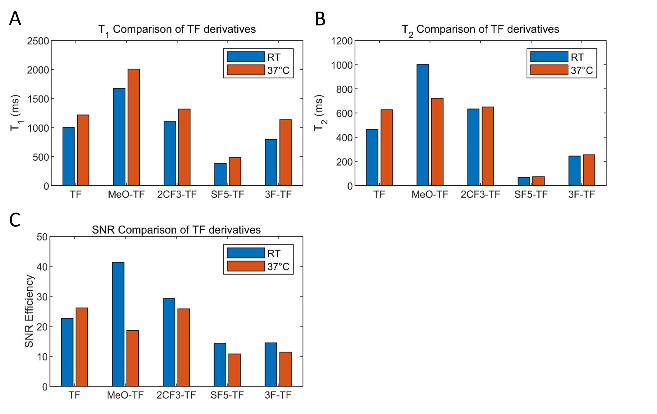
TF as well as the derivatives methoxy-teriflunomide (MeO-TF) and di-trifluormethyl-teriflunomide (2CF3-TF) showed a single 19F peak spectrum (Fig.1A-C). Penta-fluoro-sulfonyl-teriflunomide (SF5-TF) revealed two narrow major and three minor peaks (Fig.1D). Trifluoro-teriflunomide (3F-TF) provided one major and one minor peak (Fig.1E). The assessment of the T1 relaxation times yielded longer T1 values for MeO-TF and 2CF3-TF derivatives but shorter T1 values for SF5-TF and 3F-TF derivatives versus the reference compound TF (Fig.2A). For all compounds T1 increased at 37°C versus the baseline measurement at RT. The assessment of the T2 relaxation times yielded prolonged T2 values for MeO-TF and 2CF3-TF derivatives but a T2 shortening for SF5-TF and 3F-TF derivatives, with TF being the reference. At 37°C the T2 of TF was longer while that of MeO-TF was shorter than at RT. (Fig2B). The SNR assessment showed that the SNR efficiency of MeO-TF and 2CF3-TF was superior to the SNR efficiency of TF. SF5-TF and 3F-TF revealed an SNR efficiency inferior to the SNR efficiency of TF. The T2 shortening of SF5-TF and 3F-TF constrain the SNR efficiency of these TF derivatives. Enzyme inhibition assays (Fig.3A) were used to determine the concentration of the inhibitor, which reduces the enzyme activity by 50% (IC50). We found an increased inhibitory capacity for MeO-TF, SF5-TF and 3F-TF, which is reflected by a lower IC50 value, and a decreased inhibitory capacity for 2CF3-TF as shown by the increase in IC50 when compared to TF (Fig.3B).Discussion
Our data show that even slight chemical modifications to the compound teriflunomide resulted in substantial changes in the 19F NMR characteristics of the derivatives. Changes in T1 and T2 particularly for MeO-TF and 2CF3-TF resulted in enhanced 19F MR sensitivities and SNR efficiencies versus TF. Importantly, the DHODH inhibitory activity (as reflected by the IC50 values) was for the most part maintained or even increased following the chemical modifications. Only one compound (2CF3-TF) showed decreased activity.Conclusions
This study demonstrated the opportunity and feasibility of chemically modified teriflunomide derivatives to enhance SNR while preserving or even improving its biological function. This is a first step toward 19F MR drug imaging with improved SNR and informing drug treatments for patient tailored therapies. Future studies are warranted to correlate the 19F MR detectability of the compounds with their biological activity with the goal to select favorable compounds for in vivo studies.Acknowledgements
This work was supported by funding from the Germany Research Council (DFG WA2804).References
1 Reid, D. G. & Murphy, P. S. Fluorine magnetic resonance in vivo: a powerful tool in the study of drug distribution and metabolism. Drug discovery today 13, 473-480, doi:10.1016/j.drudis.2007.12.011 (2008).
2 Waiczies, H. et al. Visualizing brain inflammation with a shingled-leg radio-frequency head probe for 19F/1H MRI. Scientific reports 3, 1280, doi:10.1038/srep01280 (2013).
3 Oh, J. & O'Connor, P. W. Teriflunomide. Neurology. Clinical practice 3, 254-260, doi:10.1212/CPJ.0b013e318296f299 (2013).
4 Faber, C. & Schmid, F. in Fluorine Magnetic Resonance Imaging (ed E. T. Ahrens, Flögel U.) 3-27 (Pan Stanford Publishing, 2016).
5 Ladds, M. et al. A DHODH inhibitor increases p53 synthesis and enhances tumor cell killing by p53 degradation blockage. Nature communications 9, 1107, doi:10.1038/s41467-018-03441-3 (2018).
6 Sainas, S. et al. Design, synthesis, biological evaluation and X-ray structural studies of potent human dihydroorotate dehydrogenase inhibitors based on hydroxylated azole scaffolds. European Journal of Medicinal Chemistry 129, 287-302, doi:http://dx.doi.org/10.1016/j.ejmech.2017.02.017 (2017).
Figures