3090
Radiomic Features of the Nigrosome-1 Region of the Substantia Nigra: Using Quantitative Susceptibility Mapping to Assist in the Diagnosis of Idiopathic Parkinson’s Disease1Ruijin Hospital,school of medicine, Shanghai Jiaotong University, Shanghai, China, 2School of Biomedical Engineering, Shanghai Jiaotong University, Shanghai, China, 3Radiology, Wayne State University, Detroit, MI, United States
Synopsis
There has been a major effort to study iron deposition in the substantia nigra (SN) because of its relationship to depigmentation of iron in the nigrosome-1 area. Recently, the swallow tail sign (STS) has been introduced as a new biomarker for idiopathic Parkinson’s disease (IPD). In this work, we analyzed the STS region of the SN based on quantitative susceptibility mapping (QSM) via a support vector machine (SVM) classifier and found that this radiomic approach could help to differentiate IPD patients from healthy controls.
Introduction
Imaging the nigrosome-1 from T2* weighted iron-sensitive magnetic resonance imaging (MRI) has recently emerged as a new biomarker for idiopathic Parkinson’s disease (IPD)1. Recognizing the nigrosome-1 has been possible thanks to the presence of high iron signal surrounding it that produces what is referred to as the swallow tail sign (STS)2. The loss of the STS is thought to be due to the increase in iron content subsequent to the depigmentation of the neuromelanin in the nigrosome-1 territory. However, consistent recognition of the STS among reviewers has been difficult due to individual differences in the shape of the nigrosome-1 territory and to the choice of imaging parameters such as: scanning plane, resolution, signal-to-noise (SNR) and echo time3,4. Radiomics might have the potential to overcome these shortcomings. Therefore, we chose to explore whether radiomic features of the iron content in the SN based on QSM data could help to differentiate IPD patients from healthy controls (HCs).
Methods
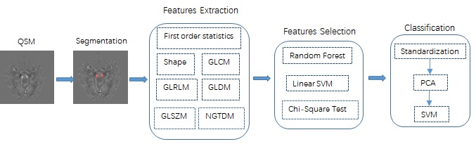
Three-dimensional multiecho gradient-recalled echo (GRE) images (0.86×0.86×1.00 mm3) were obtained at 3.0 T for QSM in 87 PD patients and 77 HCs. Regions of interest (ROIs) of the SN below the red nucleus were manually drawn on both sides, and subsequently, volumes of interest (VOIs) were segmented (these ROIs included four 1mm slices). Then, 105 radiomic features (including 18 histogram features, 13 shape features and 74 texture features) of the bilateral VOIs in the two groups were extracted using the pyradiomics tool. Forty 40 features were selected from these according to the ensemble feature selection method which combined random forest, linear support vector machine and chi-square test. The selected features were utilized to distinguish IPD patients from HC using 10 rounds of a 3-fold cross-validation support vector machine (SVM) classifier (Figure 1). Finally, the selected features were analyzed using an unpaired t-test.Results
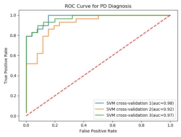
The results from SVM (Figure 2) were: area under the curve (AUC): 0.95±0.02; accuracy: 0.86±0.04; sensitivity: 0.87±0.06 and specificity: 0.84±0.09. Five features were selected to show the detailed differences between IPD patients and HCs: 10 Percentile (0.023±0.007 V.S 0.015±0.009, p<0.01 ), Median (0.076±0.016 V.S 0.066±0.015, p<0.01), and Small Area Low Gray Level Emphasis (0.277±0.119 V.S 0.242±0.109, p<0.01) of VOIs in IPD patients were higher than those of VOIs in HCs, respectively; while, Long Run Low Gray Level Emphasis (0.420±0.133 V.S 0.546±0.312, p<0.01) and Gray Level Non-Uniformity (5.769±2.442, 7.583±2.707, p<0.01) of VOIs in IPD patients were lower than those in HCs.Discussion and Conclusion
Our preliminary results showed that radiomic features of the nigrosome-1 containing region of the SN were different between IPD patients and HCs. The traditional machine learning method of SVM on nigrosome-1 containing part of SN was able to differentiate IPD from HC with a relatively high diagnostic sensitivity and specificity as in a meta-analysis reported based on visualizing the STS at 3.0 T5, though the VOIs of the STS confined regions were not delineated. The higher values of the first order (10 percentile and median) and Small Area Low Gray Level Emphasis reflected more iron content in nigrosome-1 containing region of SN in IPD patients; while, the lower values of Long Run Low Gray Level Emphasis and Gray Level Non-Uniformity reflected more uniform of the nigrosome-1 containing part of SN due to depigmentation of the neuromelanin in IPD patients, which was in consistence with the loss of STS in IPD. Therefore, the Radiomic features could help to avoid the shortcomings of subjective visualizing on recognizing this sign and, thus, could be an objective and time saving tool assisting in the diagnosis of IPD. In conclusion, radiomic features of the SN based on QSM could be useful in the diagnosis of IPD and could serve as a surrogate marker for the STS.Acknowledgements
Thanks Dr. Hongmin Xu, Yan Li and Zhijia Jin for their help in scanning and collecting data.References
1. Noh Y, Sung YH, Lee J, et al. Nigrosome 1 Detection at 3T MRI for the Diagnosis of Early-Stage Idiopathic Parkinson Disease: Assessment of Diagnostic Accuracy and Agreement on Imaging Asymmetry and Clinical Laterality. AJNR Am J Neuroradiol. 2015;36(11):2010-2016.
2. Schwarz ST, Afzal M, Morgan PS, et al. The 'swallow tail' appearance of the healthy nigrosome - a new accurate test of Parkinson's disease: a case-control and retrospective cross-sectional MRI study at 3T. PLoS One. 2014;9(4):e93814.
3. Kim EY, Sung YH, Shin HG, et al. Diagnosis of Early-Stage Idiopathic Parkinson's Disease Using High-Resolution Quantitative Susceptibility Mapping Combined with Histogram Analysis in the Substantia Nigra at 3 T. J Clin Neurol. 2018;14(1):90-97.
4. Schmidt MA, Engelhorn T, Marxreiter F, et al. Ultra high-field SWI of the substantia nigra at 7T: reliability and consistency of the swallow-tail sign. BMC Neurol. 2017;17(1):194.
5. Mahlknecht P, Krismer F, Poewe W, et al. Meta-analysis of dorsolateral nigral hyperintensity on magnetic resonance imaging as a marker for Parkinson's disease. Mov Disord. 2017;32(4):619-623.
Figures