3089
Classification of Parkinson‘s disease with diffusion magnetic resonance imaging by machine learning methods1Radiology, Union Hospital, Tongji Medical College, Huazhong University of Science and Technology, Wuhan, China
Synopsis
The diagnosis of PD is mainly based on clinical features and does not rely on imaging biomarkers. Neuroimaging studies help us better understand the pathophysiology and symptoms of PD. The aim of this study is to investigate the feasibility and performance of the PD classification using diffusion MRI of different machine learning methods.
Purpose
To investigate the feasibility and performance of the PD classification using diffusion MRI of different machine learning methods 1-2.Methods
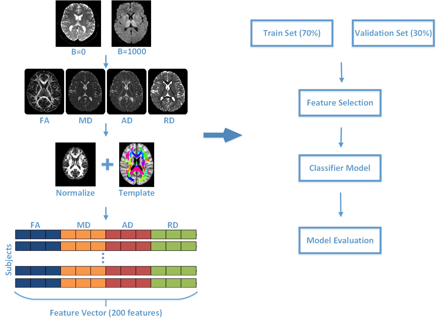
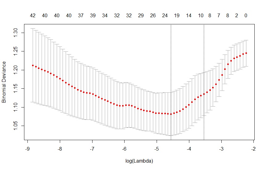
A total of 285 PD patients and 135 healthy controls were included and underwent diffusion tensor imaging scans. DTI images were processed with the FSL toolbox to get the parameters such as fractional anisotropy (FA), mean diffusivity (MD), axial diffusivity (AD), radial diffusivity (RD). Values of each brain areas in stereotaxic white matter atlas were extracted by the ICBM template as the raw features for subsequent machine learning based classifiers 3. All of the PD patients and healthy controls were separated to training data sets and testing data sets. Feature optimization was performed by the LASSO and principal component analysis. And both the filter- and wrapper-based feature selection models were used to get the final features used in the last classifier. The k-nearest neighbor algorithm (KNN), naïve Bayes algorithm and support vector machine (SVM) models were trained on the training data sets and the performance of these models were evaluated by the testing data sets.Results
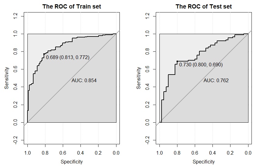
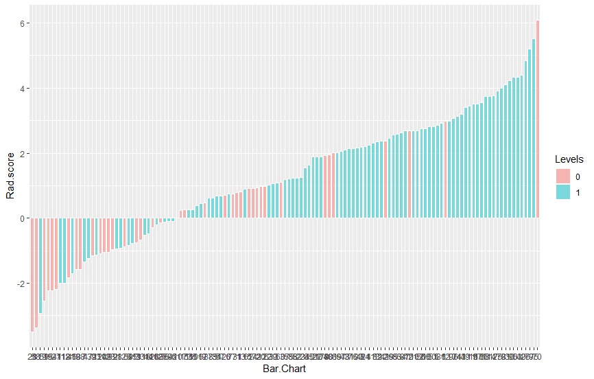
Compared to healthy control subjects, PD patients showed significantly alteration of white matters in wide spread brain regions. The machine learning analyses showed that the SVM model performs best than KNN and naïve Bayes algorithm with an overall accuracy of 86.7%, sensitivity of 83.3% and specificity of 89.0% when discriminated PD patients from healthy controls.Conclusion
The combination of DTI and machine learning methods is a promising method to discriminate PD patients from healthy controls. These findings may be useful for future incorporation of DTI and machine learning methods for disease classification, or as a useful tool for clinical diagnosis by diffusion MRI. DTI-based machine learning methods can help to better PD diagnosis.Acknowledgements
No acknowledgement found.References
1, Lanskey, J.H., et al., Can neuroimaging predict dementia in Parkinson’s disease? Brain, 2018.
2, de Vos, F., et al., A comprehensive analysis of resting state fMRI measures to classify individual patients with Alzheimer's disease. NeuroImage, 2018. 167: p. 62-72.
3, Atkinson-Clement, C., et al., Diffusion tensor imaging in Parkinson's disease: Review and meta-analysis. NeuroImage: Clinical, 2017. 16: p. 98-110.
Figures