3088
Prediction of Chronological Age from Intracranial Time-of-Flight Magnetic Resonance Angiography Images by Deep Convolutional Neural Network1Seoul National University Hospital, Seoul, Korea, Republic of
Synopsis
Brain-predicted age may be used as a potential biomarker of brain aging, and there also may be features related to cerebrovascular aging, such as decline of visualization of the arteries, or tortuosity. Therefore, the purpose of this study was to investigate whether there are learnable features by a deep convolutional neural network in the maximum intensity projection images of time-of-flight magnetic resonance angiography that might be associated with cerebrovascular aging.
INTRODUCTION
Brain-predicted age may be used as a potential biomarker of brain aging,1 and there also may be features related to cerebrovascular age. Previous studies reported the age-related features of intracranial MR angiography, such as decline of visualization of the arteries,2 or tortuosity.3 Although a prior study demonstrated correlation of age and 3D volume data of time-of-flight MRA images,4 we postulated that 3 orthogonal maximum intensity projection (MIP) images also have information of age-related cerebrovascular features. In addition, 3D volume data of time-of-flight MRAs are heavily T1-weighted and may contain structural information as well that is not related to cerebrovascular aging, if background suppression was incomplete. Therefore, the purpose of this study was to investigate whether there are learnable features by a CNN in the MIP images of time-of-flight MRA that might be associated with cerebrovascular aging.METHODS
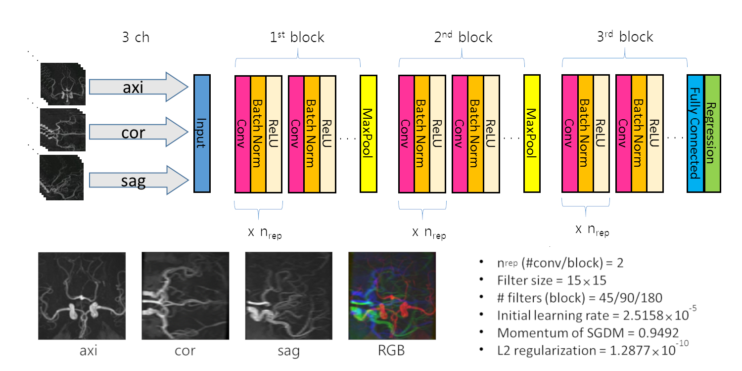
The study was approved by IRB. Two datasets were used in our study; there were 486 MRA data downloaded from the Information eXtraction from Images (IXI) repository,5 and another 500 MRAs from healthy patients who visited Health Promotion Center of our institution. Our institution’s MRA data were acquired for routine check-up without evidence of brain or cerebrovascular disease, and investigators reviewed the formal report to exclude scans with abnormal findings. The mean age of pooled dataset was 52.0 years (range; 19-88 years). The MRI scans in our institution were performed by using various scanners and main magnetic field strength; GE 3.0T (n=231), Siemens 3.0T (n=13), Philips 3.0T (n=13), GE 1.5T (n=200) and Siemens 1.5T (n=43). The data were divided randomly into a training (n=886) and a test (n=100) sets. Each MRA volume data was augmented by a factor of 10 by random scaling (0.9-1.1 times), translation along a random vector (±5 pixels), and rotation (±7.5 degree) around a randomly oriented axis in a row. Then, MIP was performed along the axial, coronal and sagittal directions. All MIP images were cropped and resampled into 115×115. Thus, the input data consist of 3 orthogonal plane MIP images from each MRA volume data; a 2.5D approach as previously described (Figure 1).6 A CNN was designed and Bayesian-optimized in Matlab (Mathworks, Inc.) (Maximum epoch=50 and mini-batch size=32).7 For the training of the CNN a stochastic gradient descent with momentum algorithm was used. The ages of the augmented data in the test set were individually predicted by the CNN, and the mean predicted age over the 10 augmented images from the same source image was used in the data analysis. The performance of CNN was evaluated by calculating the mean absolute error (MAE) and the Pearson’s correlation coefficient (r) between the ground truth and the CNN-predicted brain ages. Finally, we trained two CNNs on the IXI dataset and our institution’s dataset separately using the same hyperparameters used for the combined dataset, and cross-validated.RESULTS
The optimized CNN is shown in Figure 1. The MAE between the MRA-predicted age and the ground-truth age was 8.40 years. The r was 0.79 (Figure 2). For the two CNNs trained on the IXI dataset and our own dataset separately, the MAE and r were 19.79 and 0.09 for the former (training on the IXI dataset (n=436/50 for train/validation) and test on our institution’s dataset (n=500)), and 15.41 and 0.22 for the latter (training on our institution’s dataset (n=450/50 for train/validation) and test on the IXI dataset (n=486))(Figure 3).DISCUSSION
The MAE and r need to be further improved with more training data and better CNN design and architecture. Nonetheless, our preliminary results may support that MIP images of brain MRA possesses age-associated features learnable by a CNN, and that the features might be related to cerebrovascular aging. The MIP images are sparse, and consequently, appear to require a relatively large convolution filter size (15×15). In our study, large MAE were obtained compared to those in the published study,1 which might be caused by the heterogeneity of the images scanned by various scanners and scan parameters. Another cause might be related to the different populations (European vs. Asian). Therefore, datasets from multiple institutions or populations were not readily applicable directly to the CNNs trained on different datasets. Elaborated data standardization and external validation are mandatory. In addition, investigations on the clinical significance of the results should also be warranted, including the analysis of the differences between the brain-predicted and the chronological ages in specific cerebrovascular disease groups.CONCLUSION
The MAE and correlation coefficient obtained in our study indicate that there might be learnable features by CNN in the MIP images of MRA in association with cerebrovascular aging.Acknowledgements
No acknowledgement found.References
1. Cole JH, Poudel RPK, Tsagkrasoulis D, et al. Predicting brain age with deep learning from raw imaging data results in a reliable and heritable biomarker. Neuroimage. 2017;163:115-124.
2. Kajiya Y, Kajiya Y, Nakajo M. Age-related changes in cerebral MR angiography. J Neurol Sci. 1997 Feb 12;145(2):195-203.
3. Bullitt E, Zeng D, Mortamet B, et al. The effects of healthy aging on intracerebral blood vessels visualized by magnetic resonance angiography. Neurobiol Aging. 2010 Feb;31(2):290-300.
4. Nam Y, Lee J, Kim DH, et al. Predicting the age from time of flight MR angiography using 3D convolutional neural network. Proc. Intl. Soc. Mag. Reson. Med. 26 (2018), 2097.
5. https://brain-development.org/ixi-dataset/
6. Roth HR, Lu L, Seff A, et al. A new 2.5D representation for lymph node detection using random sets of deep convolutional neural network observations. Med Image Comput Comput Assist Interv. 2014;17(Pt 1):520-527.
7. Snoek J, Larochelle H, Adams RP. Practical bayesian optimization of machine learning algorithms. Adv Neural Inf Process Syst. 2012; 2951-2959.
Figures