3087
Automated machine learning classification of first-episode schizophrenia and controls using cerebral morphometric features1Huaxi Magnetic Resonance Research Center (HMRRC), Department of Radiology, West China Hospital of Sichuan University, Chengdu, China, 2College of Physical Science and Technology, Sichuan University, Chengdu, China, 3MR Collaboration, Siemens Healthcare Ltd., Shanghai, China, 4Department of Psychiatry, University of Cincinnati, Cincinnati, OH, United States
Synopsis
Discriminate the first-episode schizophrenia patient with optimal feature set and classification model identified by automated machine learning algorithm
Purpose
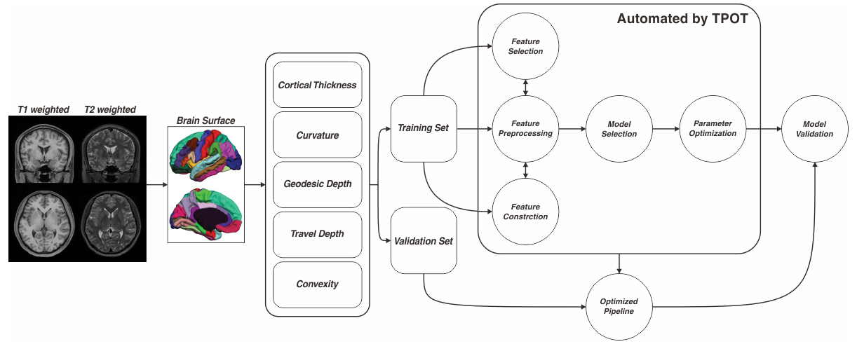
Extensive efforts have been made to identify brain changes related to schizophrenia. Unlike classical between-group comparisons, machine learning can identify subtle disease patterns in a single subject level, which is needed for clinical application. However, previous machine learning studies have two principal limitations. First, most were performed in chronic patients in which long-term illness duration and medication may alter brain morphology and thus have influenced the results. Second, most studies only utilize T1-weighted images. Recent studies have indicated that analysis with additional T2 weighted images can significantly improve the accuracy of brain surface reconstruction. The current prospective study aims to identify the optimal feature set and machine learning pipeline to discriminate between patients with first episode schizophrenia and healthy controls by using additional high resolution T2 weighted images.Methods
Fifty-four first-episode schizophrenia patients (30 male, 24 female, aged 18 to 25 years, and mean age 22.6 years) were included in this study. Confirmation of diagnosis was determined by clinical psychiatrists using the Structured Clinical Interview for DSM-IV (SCID). No patient had received any psychiatric medication before their MRI scan. Illness duration of all patients was less than 2 years. Forty-nine demographically-matched controls were recruited from the local community via poster advertisements. High resolution T1 weighted anatomical images were acquired on a clinical 3T scanner with a 32-channel phase array head coil using a MPRAGE sequence (TR=2400ms, TE=2ms, TI=1000ms, Flip angle=8, 0.8mm isotropic resolution). High resolution T2 weighted images were acquired using a 3D SPACE (TR=3200ms, TE=565ms, 0.8mm isotropic resolution) sequence.
Both T1 and T2 weighted anatomical images were processed with Freesurfer's recon-all processing pipeline with the Desikan-Killiany-Tourville (DKT) atlas to generate labeled brains. The labeled brains were then fed into the open source shape analysis software Mindboggle1. Surface meshes were generated from each segmented region. The following 5 categories of morphometric features were computed for each cortical mesh: (1) Mean cortical thickness; (2) Mean curvature; (3) Mean geodesic depth; (4) Mean travel depth; (5) Mean convexity.
A genetic algorithm based automated machine learning algorithm implemented in the Tree-based Pipeline Optimization Tool (TPOT)2 was used to construct the optimal classification pipelines. To alleviate the high dimension/low sample size problem, five categories of features were fed into machine learning pipelines, respectively. A hold-out scheme is used to evaluate each identified model. Seventy-five percent of all involved subjects were randomly selected to form the training cohort; the rest were treated as a testing cohort (Figure 1). Metrics including sensitivity, specificity, accuracy, kappa score, and area under the ROC curve (AUC) were used to assess each model.
Results
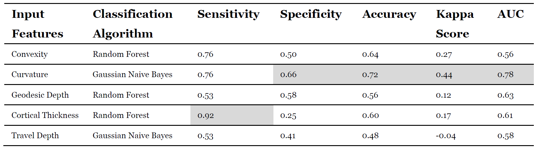
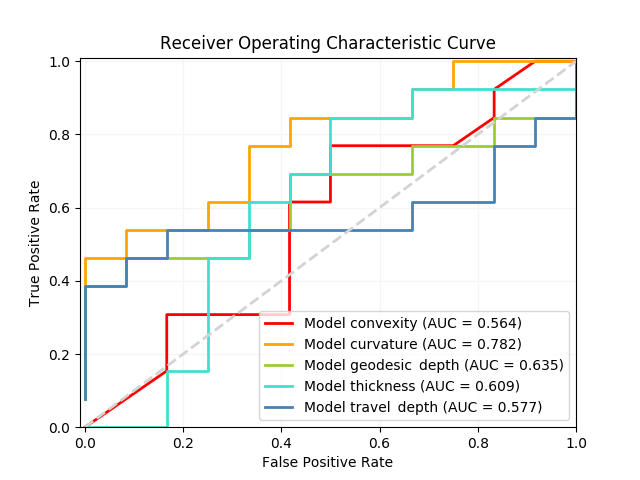
A total of 315 features (63 features for each category) representing cerebral morphometry were extracted from each participant. The performance of each automated identified pipeline was validated using the independent validation set. The identified models and performance are listed in Figure 2. ROC curves were also plotted to illustrate diagnostic performance of each model (Figure 3).Discussion
Applying machine learning techniques to MRI data has achieved impressive successes in the field of psychiatric research. However, identification of optimal machine learning pipelines for a given problem is not an easy task as there are dozens of choices to make in the steps of data preprocessing, model and feature selection, and hyperparameter tuning. The current study exploited a recently proposed automated machine learning algorithm to search the optimal model to discriminate patients with first-episode schizophrenia through cerebral morphometric features. The Gaussian Naive Bayes classifier with curvature features achieved the best performance (AUC=0.78), while the random forest classifier with cortical thickness achieved highest sensitivity (0.92) but lowest specify (0.25). The current results confirm the aberrant cortical morphology in patients with first episode schizophrenia at the level of individual patients. In addition, the current results indicate that curvature, a measure of gyrification, has more contribution to gray matter volume changes than cortical thinning3.Conclusion
Our results demonstrate that schizophrenia patients with moderate effectiveness can be identified at an early stage of illness based on cortical curvature. However, the robustness of this metric as an imaging biomarker needs further clinical validation.Acknowledgements
No acknowledgement found.References
1. Klein A, Ghosh SS, Bao FS, Giard J, Häme Y, Stavsky E, et al. Mindboggling morphometry of human brains. Schneidman D, editor. PLOS Comput Biol. 2017;13: e1005350.
2. Olson RS, Bartley N, Urbanowicz RJ, Moore JH. Evaluation of a Tree-based Pipeline Optimization Tool for Automating Data Science. Proc 2016 Genet Evol Comput Conf - GECCO ’16. New York, New York, USA: ACM Press; 2016; 485–492.
3. Kong L, Herold CJ, Zöllner F, Salat DH, Lässer MM, Schmid LA, et al. Comparison of grey matter volume and thickness for analysing cortical changes in chronic schizophrenia: A matter of surface area, grey/white matter intensity contrast, and curvature. Psychiatry Res - Neuroimaging. Elsevier; 2015;231: 176–183.
Figures