3083
Improved classification of paediatric brain tumours through whole spectra from in vivo magnetic resonance spectroscopy1Institute of Cancer and Genomic Sciences, University of Birmingham, Birmingham, United Kingdom, 2Birmingham Children’s Hospital NHS Foundation Trust, Birmingham, United Kingdom
Synopsis
Single voxel magnetic resonance spectroscopy (SVS) is a non-invasive technique that can be used to probe metabolic activity in tumours. Previous studies have used metabolite concentrations to classify paediatric brain tumours from good quality data. However, the use of in vivo MRS whole spectra and wavelet de-noising for paediatric brain tumours have been rarely reported. In this study, we investigated the performance of spectra in classifying paediatric brain tumours by employing wavelet-based de-noising, and found significantly reduced error rate of classification based on the whole spectra, compared to that from metabolite concentrations and fits.
Introduction
Single voxel magnetic resonance spectroscopy (SVS) is a non-invasive technique that can be used to probe metabolic activity in tumours. Previous studies have used metabolite concentrations to classify paediatric brain tumours, achieving classification accuracies between 93%-95%1. However, the use of in vivo MRS whole spectra for paediatric brain tumours have been rarely reported. Previously, we investigated the use of wavelets to improve data quality, and our results showed reduced bias of metabolite concentration estimation2. Other studies showed that complete tumour spectra were able to perform a classification according to the highest estimated tissue proportion, based on the adult brain tumours glioblastoma multiforme, low-grade gliomas and meningiomas3. In this study, we aim to investigate the performance of spectra in classifying paediatric brain tumours by employing wavelet-based denoising, and further compared to that based on metabolite concentrations and fits.Materials and methods
Clinical MRS was acquired using a Siemens 1.5T scanner with the SVS Point-RESolved Spectroscopy (PRESS, TE=30ms, TR=1500ms) at Birmingham Children’s Hospital. Wavelet bases were selected based on the maximum SNR that can be achieved from all available mother wavelets including daubechies, biorthogonal, coiflets, and symlets. De-noising was performed using wavelet transform with 2nd level of decomposition and level-dependent thresholding using Matlab (The Mathworks, MA). TARQUIN 4.3.11 was used to quantify MRS, and providing fits and metabolite concentrations for analysis, cases with failed qualification by quality control matrix in TARQUIN software have been excluded. In total, sixty-eight cases were studied, 7 infratentorial ependymomas (EP), 30 medulloblastomas (MB) and 31 pilocytic astrocytomas (PA).
Spectra and fits were normalised prior to feature extraction. Classification was achieved through the combination of principal component analysis (PCA) for dimension reduction and linear discriminant analysis (LDA) for clustering on the metabolite concentrations, fits or spectra individually. In both spectra and fit analysis, metabolites with a chemical shift between 0.2 and 4ppm were used for classification. All metabolite components available in TARQUIN had been included in fits-based classification. The number of principal components used in LDA was set to obtain 95% of the cumulative variance. Classification accuracy was obtained by re-substitution, leave one out cross validation, bootstrap .632 and .632 plus cross validation.
Results
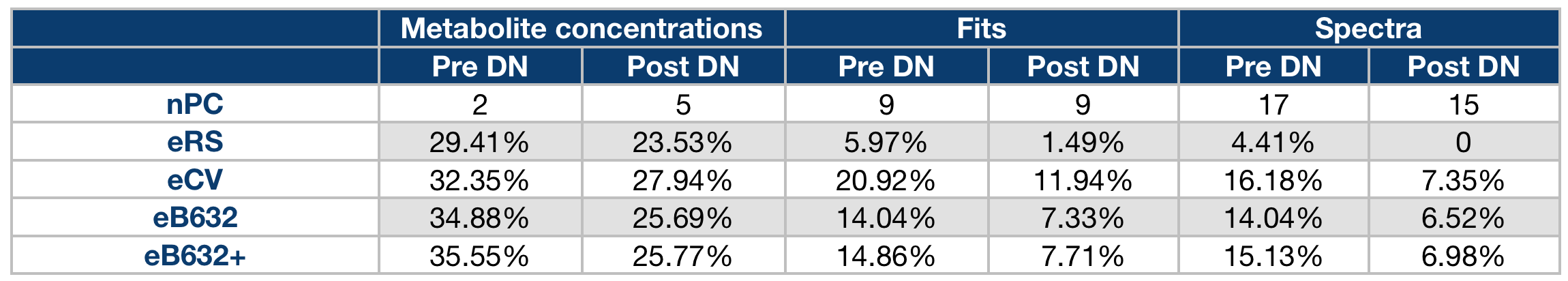
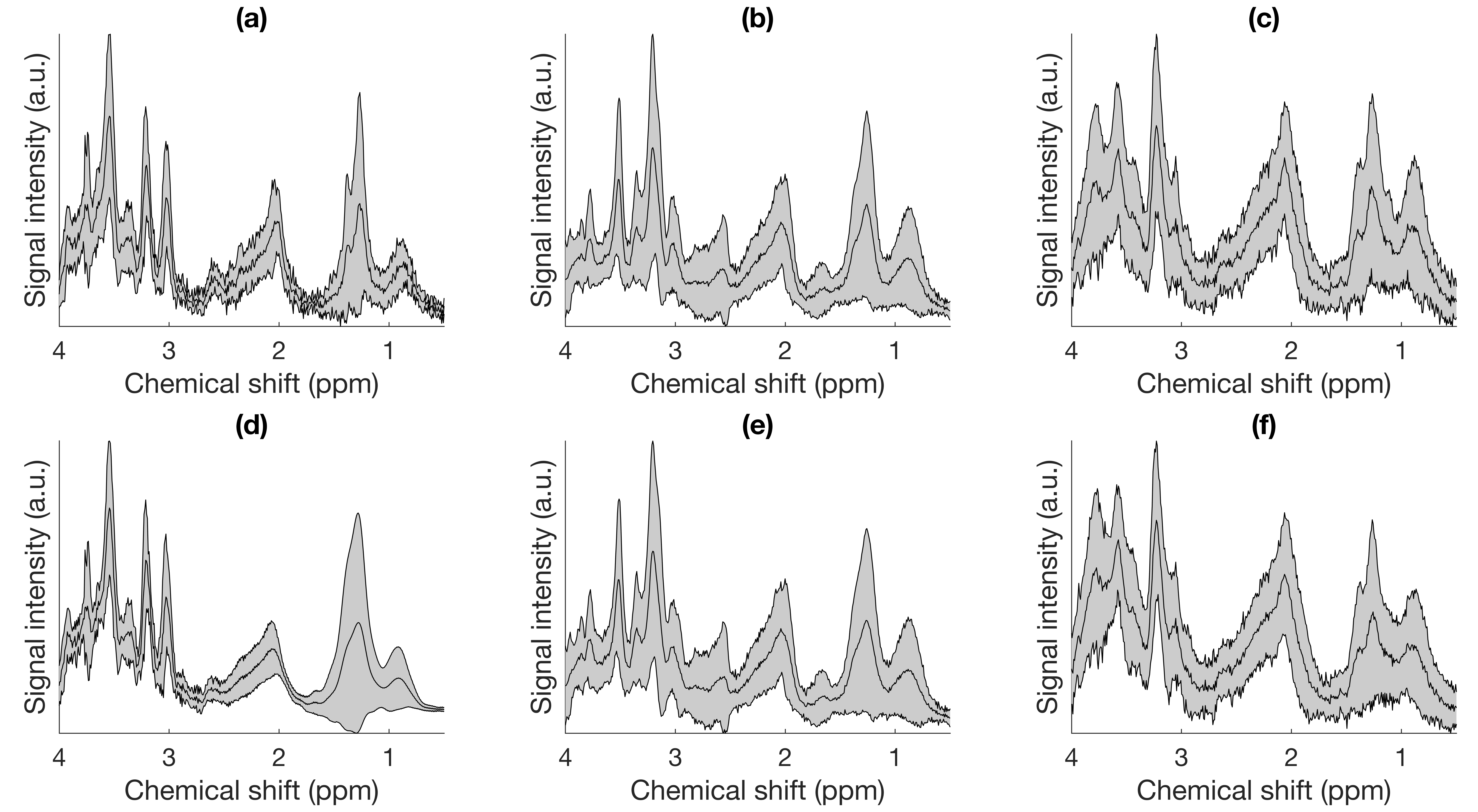
Figure 1 shows the average explained variance as derived from principle components (PC). Sixteen PCs were employed for classification since 95% of the total cumulative variance can be explained from pre-DN MRS, and fifteen PCs were employed for post-DN MRS. Figure 1 also shows improved classification for three tumours after DN based on two discriminant function scores. Figure 2 shows mean and standard deviation of spectra for the three tumours before and after DN. Table 1 shows classification accuracy explained by apparent error rate based on re-substitution and true error rate based on leave-one-out cross validation, .632 and .632 plus bootstrap cross validation.Discussion
We have used both fits and whole spectra of de-noised MRS to classify paediatric brain tumours, using a combination of PCA and LDA, and also use the pre and post de-noising MRS to observe the impact of wavelets in tumour classification. Results of principal component decomposition show more information can be used from spectra (nPre-DN=17, nPost-DN=15), compared to fits (nPre-DN=9, nPost-DN=9) and metabolite concentrations (nPre-DN=2, nPost-DN=5). Significantly decreased error rate of classification appears from metabolite concentrations to fits and then spectra, also from pre to post de-noising. Impact of wavelets were also reflected by the improved distribution of three tumour groups based on the discriminant function scores compared to that from the unprocessed MRS. Current results of paediatric brain tumour classification demonstrate de-noised MRS as a potential method to improve classification accuracy employing by wavelets, but the spectra show missing metabolites by eye after DN, like lactate in ependymomas, which will be investigated based on CR bounds in the next step. Since ependymomas are relatively rare and hard to collect, the sample size of ependymomas is not as large as that of the other two tumour types, thus it might be challenging to classify more unknown ependymoma cases based on the current six samples. Further refinements are needed to ensure cases near the boundary can be better classified after de-noising.Acknowledgements
No acknowledgement found.References
- Davies NP, Wilson M, Harris LM et al. Identification and characterisation of childhood cerebellar tumours by in vivo proton MRS. NMR Biomed. 2008;21:908-918.
- Zhao D, Sun Y, Peet AC et al. Improving MRS classification of children’s brain tumours through wavelet de-noising. Proceedings the 24th British Chapter of ISMRM Annual Meeting. Oxford, England, 24-26 Sep 2018.
- Raschke F, Fuster-Garcia E, Opstad KS, Howe FA. Classification of single-voxel 1H spectra of brain tumours using LCModel. NMR Biomed. 2012;25:322-331.
Figures