3079
A Transfer Learning-based Radiomics Model for Prediction MGMT Promotor Methylation Status in Glioblastoma Multiforme1Department of Radiology, Guangzhou First People's Hospital, School of Medicine, South China University of Technology, Guangzhou, China, 2Philips Healthcare, Guangzhou, China, 3Department of Radiology, Guangdong General Hospital, Guangzhou, China
Synopsis
Glioblastoma multiforme (GBM) is the most common malignant brain tumor. MGMT promoter methylation is associated with beneficial chemotherapy. We extract deep features from a pre-trained deep neural network model via transfer learning and generate an effective feature vector model together with radiomics features for an optimal pretreatment prediction of MGMT promoter methylation status. The deep feature set achieved the higher predictive accuracy of 0.86 and 0.70 for validation and test group comparing to handcrafted radiomics feature and combined feature sets. The deep feature model may serve as a potential imaging biomarker for pretreatment prediction of MGMT methylation in GBM.
Introduction
Glioblastoma multiforme(GBM) is the most common malignant brain tumor. Promoter methylation of the DNA repair gene O6-methylguanine-DNA methyltransferase(MGMT) is associated with beneficial chemotherapy1. This study aims to extract deep features from a pre-trained deep neural network model via transfer learning, and generate an effective feature vector model together with radiomics features for an optimal pretreatment prediction of MGMT promoter methylation status in GBM.Materials and Methods
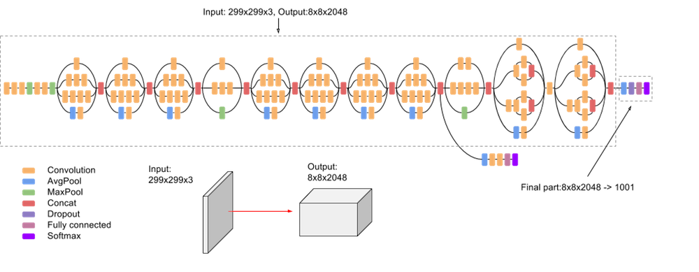
134 GBM patients (allocated randomly to a train, validation and test set with a ratio of 8:1:1) with known MGMT status. A region of interest(ROI) of the tumor was manually delineated on preoperative post-contrast T1WI for each patient. Slice thickness, image matrix and signal intensities range of all images were normalized to 3mm, 299 x 299 and [0, 255], respectively. Then 1412 handcrafted radiomics features including 1st order, shape-based, gray level-based and wavelet-based parameters were extracted from the ROIs via pyradiomics platform. Due to the small cohort size, a transfer learning method was applied2. A pre-trained neural network model named as InceptionV3 with high recognition accuracy was used for deep feature extraction, as shown in Figure 1. The pre-trained Inception-v3 model achieves state-of-the-art accuracy for recognizing general objects with 1000 classes. The model extracts general features from input images in the first part and classifies them based on those features in the second part. All images were input into the network model with an exclusion of final fully connected layer in order to get deep feature vector for a later machine learning process. The general average pooling output with size 2048 of InceptionV3 model was defined as a deep feature3, as it represents the feature vector before pattern recognition process in deep neural network. Afterwards, all 1412 handcrafted features and 2048 deep features for each patient were normalized as z-scores. Features with zero median absolute deviation(MAD) values were discarded which were considered as non-informative, and 41 features were excluded. Then, the area under the receiver operating characteristic curves (AUC) of each feature vector were calculated with reference to MGMT outcome. Features with AUC>=0.58 were regarded as good predictor factors, and 3388 feature vectors were discarded. Additionally, the number of feature vectors was further reduced by discarding highly correlated features. If the correlation coefficient between a pair of features was >=0.90, the feature with higher AUC was kept 4. Finally, 13 image feature vectors were remained and regarded as robust, non-redundant and predictive. Among them, there were 3 deep learning features with average AUC 0.6 and 10 radiomics features with average AUC 0.59. An AUC weighted support vector machine method was applied for MGMT pattern recognition5. The AUC represents the probability that a randomly chosen positive example is correctly ranked with greater suspicion than a randomly chosen negative example. We formed radiomics feature vectors according to their AUC values, during the SVM validation test, the weight of each feature was fine tuned. After validation, three sets of feature vectors were formed: deep feature, radiomics feature and combined feature. The deep feature vectors were 0.8*Inceptionv3_2193 + 0.2*Inceptionv3_2209; The radiomics vectors were given as 0.8* radiomics_551 + 0.2*radiomics_1022; The combined feature vector was 0.8*Inceptionv3_2193 + 0.2*radiomics_551.Results
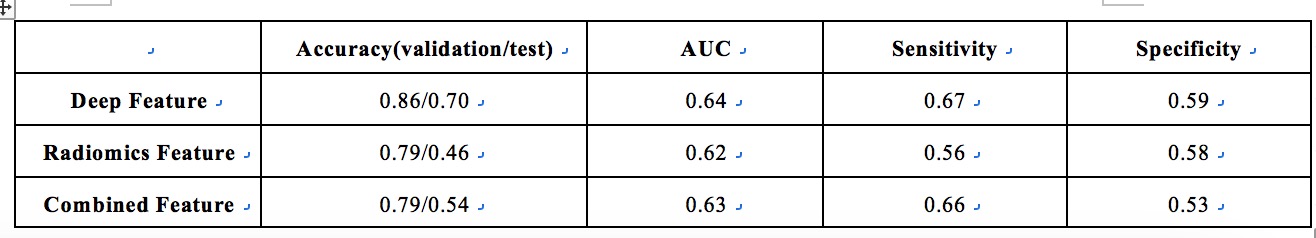
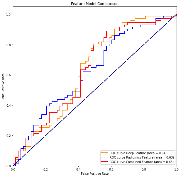
The diagnostic accuracy of deep feature set for validation and test group was 0.86 and 0.70, while that of radiomics features was 0.78 and 0.46, and combined feature set had accuracy 0.79 and 0.54, respectively. Obviously, the deep feature set demonstrated the best accuracy in predicting MGMT. The ROC result
was
shown in Figure 2 and 3, and deep feature set had the highest AUC 0.64. Its
sensitivity 0.67 and specificity 0.59 were also higher than others.Discussion
Conclusion
For a relatively small size of medical image dataset, the pre-trained deep learning network-based model combined with a well-defined support vector machine method may serve as a potential imaging biomarker for pretreatment prediction of MGMT methylation in GBM.Acknowledgements
This work was supported by the National Natural Scientific Foundation of China (grant numbers 81601469 and 81771912).References
1. Hegi ME, Diserens AC, Gorlia T, et al. Mgmt gene silencing and benefit from temozolomide in glioblastoma [J]. N Engl J Med, 2005,352(10):997-1003.
2. Lao J, Chen Y, Li ZC, et al. A deep learning-based radiomics model for prediction of survival in glioblastoma multiforme. Scientific reports. 2017;7(1):10353.
3. Christian Szegedy, Vincent Vanhoucke, Sergey Ioffe, Jonathon Shlens. Rethinking the Inception Architecture for Computer Vision arXiv:1512.00567v3 [cs.CV] 11 Dec 2015
4. Shaoyi Zhang M. Maruf Hossain Md. Rafiul Hassan James Bailey, Feature Weighted SVMs Using Receiver Operating Characteristics, International Conference on Data Mining, 2019;497-508
5. Alfredo Canziani & Eugenio Culurciello,,Adam Paszke , An analysis of deep neural network models for practical application. arXiv:1605.07678v4 [cs.CV] 14 Apr 2017
Figures