3078
Integrating Machine Learning and Image Inpainting to Predict Tumour Invasion in Glioblastoma using multi-parametric MRI1Department of Clinical Neurosciences, University of Cambridge, Cambridge, United Kingdom, 2Department of Neurosurgery, Shanghai General Hospital, Shanghai Jiao Tong University School of Medicine, Shanghai, China, 3The Centre for Mathematical Imaging in Healthcare, Department of Pure Mathematics and Mathematical Statistics, University of Cambridge, Cambridge, United Kingdom, 4Department of Radiology, University of Cambridge, Cambridge, United Kingdom
Synopsis
The multi-parametric MRI has the potential to compensate for the non-specific contrast-enhancing imaging in delineating tumor margin. The purpose of this study was to propose a method by integrating machine learning with image inpainting to predict the glioblastoma invasion using advanced multi-parametric MRI. The predictive tumor regions using this approach showed significance for patient prognosis, in a cohort containing 115 glioblastoma patients. This approach could advance the scenario of mathematical image analysis by considering both imaging features and brain structure. The predictive region may have significant clinical impact on personalized and targeted surgical treatment of patients.
Introduction
Glioblastoma is the most common primary malignant tumour in adults, characterized by poor outcomes. Evidence shows that a higher extent of resection is beneficial. However, extended resection may subject patients to higher risks. Therefore, accurately targeting is crucial for surgical planning. Multi-parametric MRI has been developed to compensate for the non-specific contrast-enhancing imaging in tumor delineation. However, effective models to integrate the multifaceted information from multiple modalities remains a challenge. Machine learning algorithms are successful in integrating multi-parametric MRI. This method, however, has the limitation of being purely driven by the feature values, without considering the brain crucial anatomical and structural information. Image inpainting is a technique to reconstruct lost or corrupted (deteriorated/blurred) information based on the image structure [1]. Therefore, the purpose of this study is to investigate whether integrating machine learning and image inpainting into multi-parametric MRI analysis could better delineate tumor invasion.Methods
Patients
This study was approved by the local institutional review board. We prospectively recruited 115 patients (mean age 59.3 years, range 22 - 76 years, 87 males) with supratentorial de novo glioblastoma. All subjects received maximal safe tumor resection and diagnosis was confirmed by pathology. After surgery, temozolomide chemoradiotherapy was performed following the Stupp protocol. The Response Assessment in Neuro-oncology criteria was used to evaluate patient response [2].
Imaging processing
All MRI sequences were pre-operatively acquired using a 3T MRI scanner. MRI sequences include T2-weighted, post-contrast T1-weighted and FLAIR images, as well as dynamic susceptibility contrast (DSC), diffusion tensor imaging (DTI) and multivoxel 2D 1H-MRS chemical shift imaging (CSI). All images were co-registered to T2W images. The mean diffusivity (MD), fractional anisotropy (FA), isotropic (p) and anisotropic (q) maps were generated from DTI. The relative cerebral blood volume (rCBV), mean transit time (MTT) and relative cerebral blood flow (rCBF) were generated from the DSC images after leakage corrected. Contrast-enhancing (CE) and non-enhancing (NE) regions of interest were manually delineated and cross-validated by three experienced researchers on the co-registered post-contrast T1 and FLAIR images.
Mathematical modelling
1. Pre-processing by neural network. Neural network was applied to build a classification function between CE and NE region,$$$u_{NN}$$$, to collect the information from the co-registered multi-parametric MRI for inpainting.
2. Tumor invasion construction by image inpainting. A predictive tumor region was generated by using a bespoke inpainting scheme. That is, the unknown region in the inpainting problem was set to be NE, whereas the classification function $$$u_{NN}$$$ was used as the inpainting fidelity function. The predicted invasion region $$$u_{t,\mathcal{B}}$$$ was then determined from the following variational problem
$$u_{t,\mathcal{B}}:=argmin\{ {\parallel u-u_t \parallel}_{L_2(Q \backslash D)}^2 + PGV_{\mathcal{B}}(u):u\in L^1(Q) \}$$
where $$$u_t:=tu_{NN}+(1-t)χ_{CE}$$$, for $$$t∈[0,1]$$$.
3. Optimal parameters by machine learning. The parameter $$$t∈[0,1]$$$ and $$$\mathcal{B} \in \mathbb{M}$$$ were used to adjust the predictive performance of the inpainted invasion region $$$u_{t,\mathcal{B}}$$$. To achieve the optimal parameter using the following bi-level training scheme [3].
Level 1: $$$(\tilde{t}, \tilde{\mathcal{B}}) = argmin\{ \mathcal{C}(t, \mathcal{B}):t\in [0,1], \mathcal{B} \in \mathbb{M} \}$$$
Level 2: $$$u_{t,\mathcal{B}}=argmin\{ {\parallel u-u_t \parallel}_{L_2(Q \backslash D)}^2 + PGV_{\mathcal{B}}(u):u\in L^1(Q) \}$$$
The function $$$ \mathcal{C}(t, \mathcal{B}) $$$ in Level 1 was the cost function constructed by using the Cho/NAA ratio calculated from CSI.
Statistical analysis
To evaluate the significance of the predictive invasive region to patient overall survival (OS) and progression-free survival (PFS), we used the Kaplan-Meier and Cox proportional hazards regression. For Cox regression, all confounders, including IDH-1 mutation, MGMT methylation, sex, age, extent of resection and contrast-enhancing tumor volume, were considered.
Results
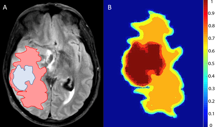
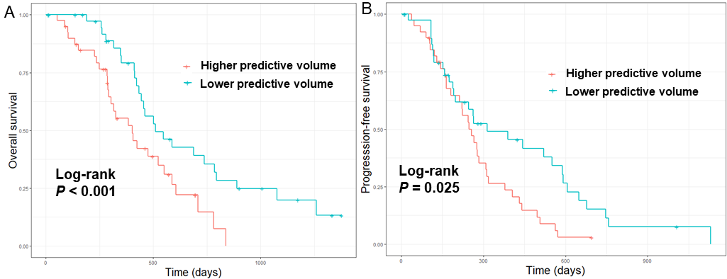
The volumes of the CE and NE regions are 53.6 ± 33.8 cm3 and 62.5 ± 44.0 cm3, respectively. The volume of the predictive regions is 31.0 ± 17.7 cm3. An example of the predictive region is shown in Figure 1. In the multivariate model, the predictive volume was significantly associated with better OS (HR = 0.97, P = 0.006). Extent of resection (HR = 2.00, P = 0.040) and tumor volume (HR = 1.04, P < 0.001) also significantly affected overall survival. Using the median ratio of the predictive volume to the CE volume, the patient subgroup with a higher ratio showed significantly better PFS (P = 0.009) and OS (P = 0.021) (Figure 2).Discussion
The proposed model considers both tumor imaging features and brain structure reflected by the multi-parametric MRI, by integrating machine learning and image inpainting. The prediction was based on the contrast-enhancing tumor core. Since the survival was only analyzed in the patients who received standard treatment, our results may suggest that the predictive regions may have better response to chemoradiotherapy, possibly due to the similar proliferative activity of this region with contrast-enhancing tumor. Further study will focus on the validation of the predictive model for treatment target.Acknowledgements
The research was supported by the National Institute for Health Research (NIHR) Brain Injury MedTech Co-operative based at Cambridge University Hospitals NHS Foundation Trust and University of Cambridge; The views expressed are those of the author(s) and not necessarily those of the NHS, the NIHR or the Department of Health and Social Care (SJP, project reference NIHR/CS/009/011); Cambridge Trust and China Scholarship Council (CL & SW); CBS acknowledges support from the Leverhulme Trust project on ‘Breaking the non-convexity barrier’, EPSRC grant Nr. EP/M00483X/1, the EPSRC Centre Nr. EP/N014588/1, the RISE projects CHiPS and NoMADS, the Cantab Capital Institute for the Mathematics of Information and the Alan Turing Institute.References
[1] Schönlieb CB. Partial Differential Equation Methods for Image Inpainting. Cambridge University Press; 2015 Oct 26.
[2] Wen PY, Macdonald DR, Reardon DA, et al. Updated Response Assessment Criteria for High-Grade Gliomas: Response Assessment in Neuro-Oncology Working Group. Journal of Clinical Oncology. 2010;28(11):1963-72.
[3] Fonseca, I., Davoli, E. and Liu, P. Adaptive image processing: first order PDE constraint regulariser and a bilevel training scheme. (submitted).
Figures