3075
Development and Validation of a Radiomics model Based on Conventional MRI for Preoperative Prediction of Gliomas with IDH1 Mutations1The First Affiliated Hospital of Dalian Medical University, Dalian, China, 2GE healthcare, Beijing, China
Synopsis
As the most common malignant tumor in the central nervous system, glioma is characterized by low progression-free survival. Studies show that abnormal expression of isocitrate dehydrogenase 1(IDH1) is closely related with the occurrence of brain tumors, especially gliomas. Evidence suggests that gliomas with mutated IDH1 have improved prognosis compared to those with wild-type IDH1.Radiomics means the high-throughput extraction of large amounts of quantitative image features from radiographic images, including segmenting tumors, building models, and then predicting and analyzing those massive feature data to assistphysicians. In this study, the IDH1 mutation was predicted by such radiomics modeling.
Objective
To evaluate the differentiation efficiency of a radiomics model based on T1WI, T2WI and contrasted T1WI MRI sequences of gliomas with and without IDH1 mutation.Materials and Methods
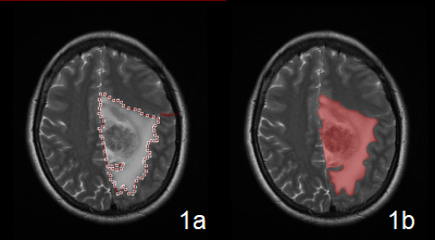
42 patients with gliomas histo-pathologically confirmed were enrolled , including 21 IDH1 mutant (IDH1-mutated; 11 males, 10 females, mean age=43.81±10.79yrs) and 21 IDH1 wild-type (IDH1-WT;10 males, 11 females, mean age=56.95±10.94yrs). The Regions of interest(ROIs) covering the entire tumor and edema were manually delineated on all axial slices using Omni-Kinetics software, and the radiomics features were automatically generated(Figure 1). All features were compared between groups. We selected features with P < 0.05 and removed redundant data. Spearman analysis was conducted to eliminate features with the correlation higher than 0.9 . Logistic regression analysis was used to establish the model for the meaningful features(Table1). Confusion matrix was used to analyze the accuracy of the model. Receiver operating characteristic (ROC) curve was plotted to assess the differential diagnostic efficiency of the features after modeling(Joint Variable(JV)).Results
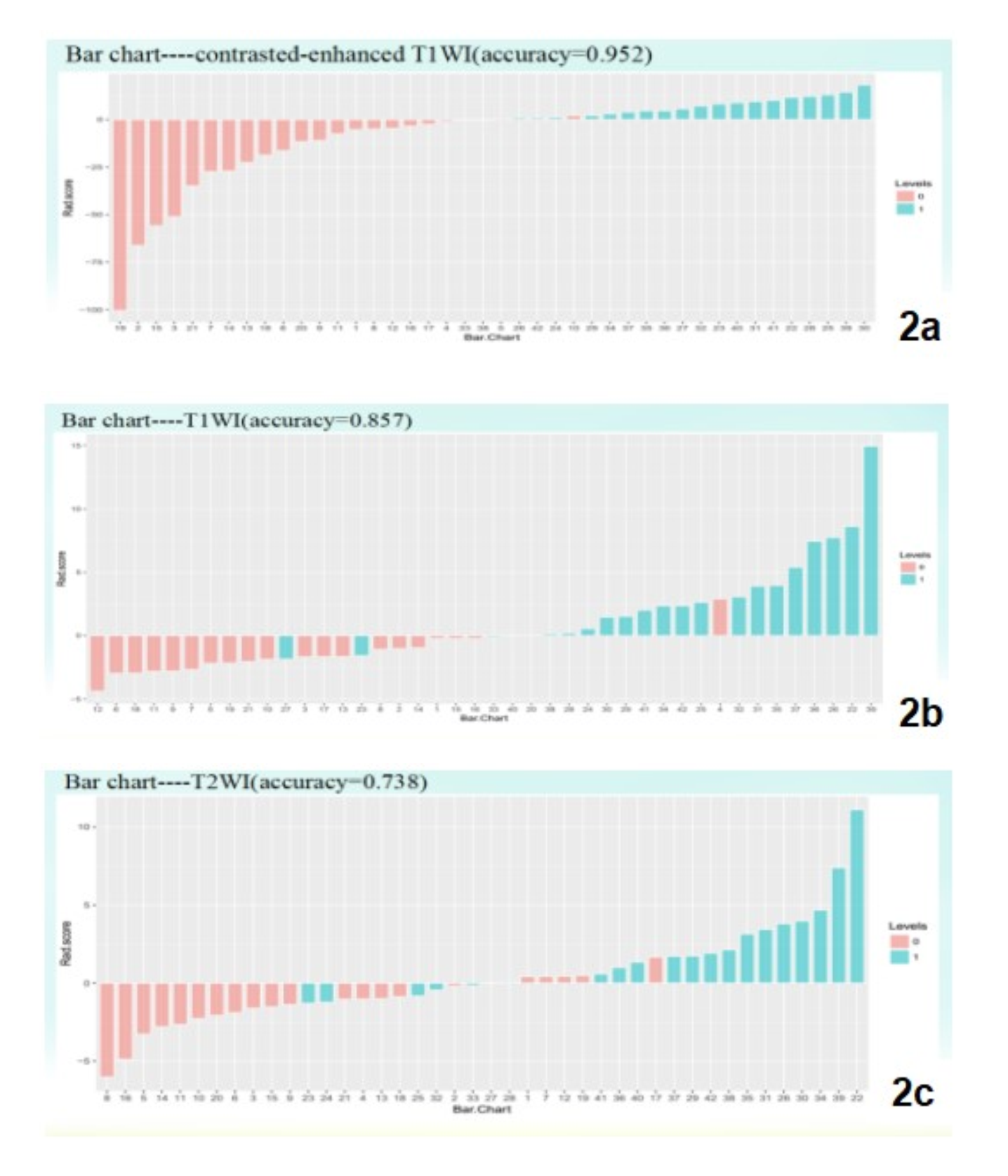
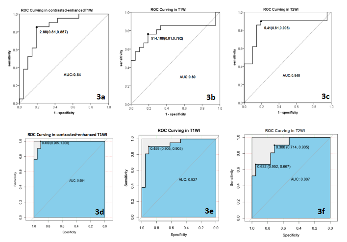
According to ROC analysis of independent variables, the IVT1WI+C(inertia) (the area under the curve (AUC) =0.844) was considered as the best feature for diagnosis(sensitivity of 85.7% and specificity of 81%). The AUC of IVT1WI(cluster prominence) for predicting IDH mutation was 0.800(specificity of 81% and sensitivity of 76.2%). IDH1-mutation was differentiated on T2WI with highest AUC(0.848) corresponding to IVT2WI (GLCM Entropy)(sensitivity=90.5%, specificity =81%). After modeling, the accuracy of contrasted T1WI, T1WI, T2WI feature modelswere 0.952, 0.857, 0.738, respectively(Figure 2). The AUC of JVT1WI+C for predicting IDH1 mutation was 0.984 (specificity of 90.5% and sensitivity of 100%). The AUC of JVT1WI for predicting IDH1 mutation was 0.927 (specificity of 90.5% and sensitivity of 90.5%). The diagnosis efficiency of JVT2WI was also satisfying (AUC=0.887, the highest specificity of 95.2%, the highest sensitivity of 90.5%)(Figure3).Discussion
Radiomics provides high throughput extraction and analyzes a large number of quantitative image features, which are used to conduct early diagnosis and construct short-term risk assessment model, and to effectively predict the development of the disease, to make a diagnosis and to trace the prognosis[1-2]. It extracts features from the region of interest that reflect the characteristics of the tumor through image segmentation, gray histogram analysis, and characteristics of the tumor shape, texture, location with its surrounding tissues[3].The study shows that the contrasted-enhanced T1WI feature model had the highest accuracy and the best diagnostic efficiency. The accuracy was up to 95.2%, and the sensitivity and specificity were up to 100%, 95.2%, respectively. The accuracy (92.7%), sensitivity (90.5%) and specificity (90.5%) of the T1WI feature model were next to the contrasted T1WI feature model. This indicates that combined radiomics features can improve diagnostic efficiency. Radiomics model has high diagnostic efficiency for IDH1 mutation, which can guide clinical treatment and prognosis evaluation.Conclusion
Radiomics model is capable for precise detection of IDH1-mutatedin gliomas. The multiple variables derived from conventional MR imaging radiomics features can improve the differentiation efficiency of gliomas with and without IDH1 mutation.Acknowledgements
No acknowledgement found.References
[1] Parson DW, Jones S, Zhang X, et al. An integrated genomic analysis ofhuman Glioblastoma multiform [J]. Science,2008,321 (5897):1807-1812.
[2] Yan H, Parsons DW, Jin G, et al. IDH1 and IDH2 mutations in gliomas[J]. N Engl J Med, 2009,360(8):765-773.
[3] Gao H, Chae O. Individual tooth segmentation from CT images using level set method with shape and intensity prior[J]. Patt Recog, 2010, 43 (7): 2406–2417.
Figures