3065
Brain connectivity and gray matter volume changes following donepezil treatment in Alzheimer’s disease1Advanced Institute of Aging Science, Chonnam National University, Gwangju, Korea, Republic of, 2Department of Radiology, Chonnam National University Medical School, Gwangju, Korea, Republic of
Synopsis
Donepezil treatment is associated with improved cognitive performance in patients with Alzheimer’s disease (AD), and its clinical effectiveness has been demonstrated. However, it has been unknown how donepezil treatment influences white matter (WM) connectivity and gray matter (GM) morphology in AD. The purpose of this study was to evaluate the thalamo-cortical white matter connectivity and GM volume after donepezil treatment in patients with AD using probabilistic tractography and voxel-based morphometry (VBM).
Introduction
The key clinical symptom of AD is a progressive deterioration of learning and memory ability which is associated with reduced acetylcholine (ACh) levels in the brain.1 Treatment with acetylcholinesterase inhibitors (AChEIs) prevents the breakdown of ACh in patients with AD and increases cholinergic transmission. To date, four AChEIs (donepezil, galantamine, rivastigmine, and tacrine) have been approved by the United States Food and Drug Administration (FDA) for treatment of the symptoms of AD by ameliorating cognitive decline by inhibiting acetylcholinesterase activity.1 Among the four drugs, donepezil is the most frequently prescribed.2,3 Donepezil has been demonstrated to inhibit acetylcholinesterase activity in the cerebral cortex, hippocampus, and striatum of the rat brain, producing increased ACh activity in the brain areas associated with cognitive function.4,5 Recently, probabilistic tractography method in diffusion weighted imaging (DWI) is becoming increasingly important in the detection of white matter integrity of an entire bundle, which allows us to estimate tract-epidemic white matter integrities by objectively assessing the tract structure.6,7 The combined use of probabilistic tractography and voxel-based morphometry may advantageously provide more valuable information regarding the thalamo-cortical white matter connectivity and GM volume after donepezil treatment in AD.Methods
Ten patients with AD (mean age = 72.4 ± 7.9 years) and 9 age-matched healthy controls (mean age = 70.7 ± 3.5 years) participated in this study. The symptom severity of AD was evaluated using the questionnaires of the mini-mental state examination (MMSE) and AD assessment scale−cognitive subscale (ADAS-Cog) before and after donepezil treatment. Ten patients with AD underwent the MR examinations before (baseline) and after (follow-up care) donepezil treatment. The treatment duration for the patients was 192.5 ± 29.6 days. The MRI and DWI were performed on a 3.0-T Magnetom Tim Trio MR Scanner (Siemens Medical Solutions, Erlangen, Germany) with an 8-channel receive head coil of birdcage type. To evaluate the thalamo-cortical WM connectivity, the regions of interest (ROIs) were divided in each individual’s T1 imaging using FreeSurfer as follows: seed region is the thalamus; target regions are the orbitofrontal cortex (OFC), medial prefrontal cortex (MPFC), lateral prefrontal cortex (LPFC), sensorimotor cortex (SMC), parietal cortex (PC), medial temporal cortex (MTC), lateral temporal cortex (LTC), and occipital cortex (OC) (Fig. 1). MRI and DWI data were post-processed using FreeSurfer and FMRIB Software Library (FSL) and Statistical Parametric Mapping (SPM8) software with the diffeomorphic anatomical registration through an exponentiated Lie algebra (DARTEL) algorithm.Results and discussion
The average MMSE scores in healthy controls (n = 9), untreated patients with AD (n = 10), and donepezil-treated patients with AD (n = 10) were 28.6 ± 1.1, 16.3 ± 4.9, and 17.6 ± 3.5, respectively, giving a significant difference in the MMSE (P = 0.031) between untreated patients and donepezil-treated patients. Average scores on the ADAS-Cog in untreated and donepezil-treated patients were 25.3 ± 8.0 and 25.1 ± 6.1, respectively (P = 0.759, NS).
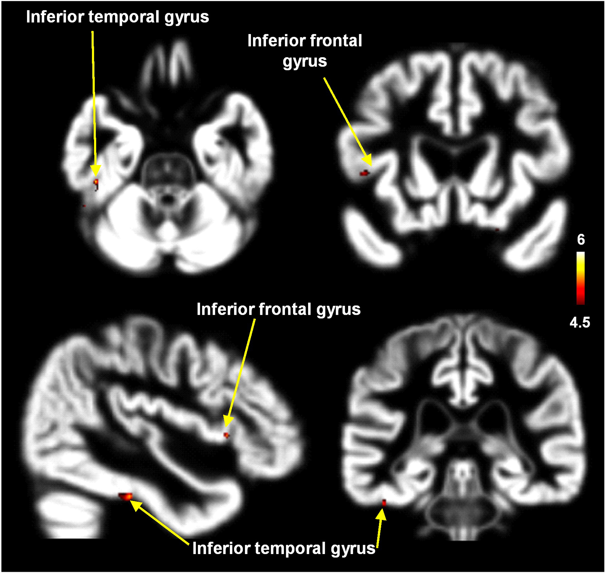
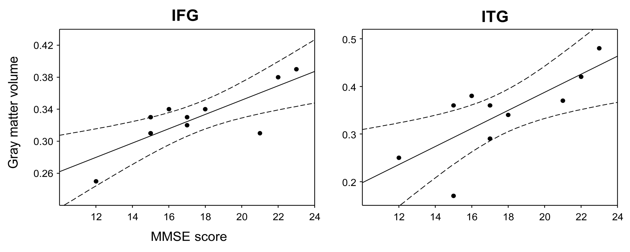
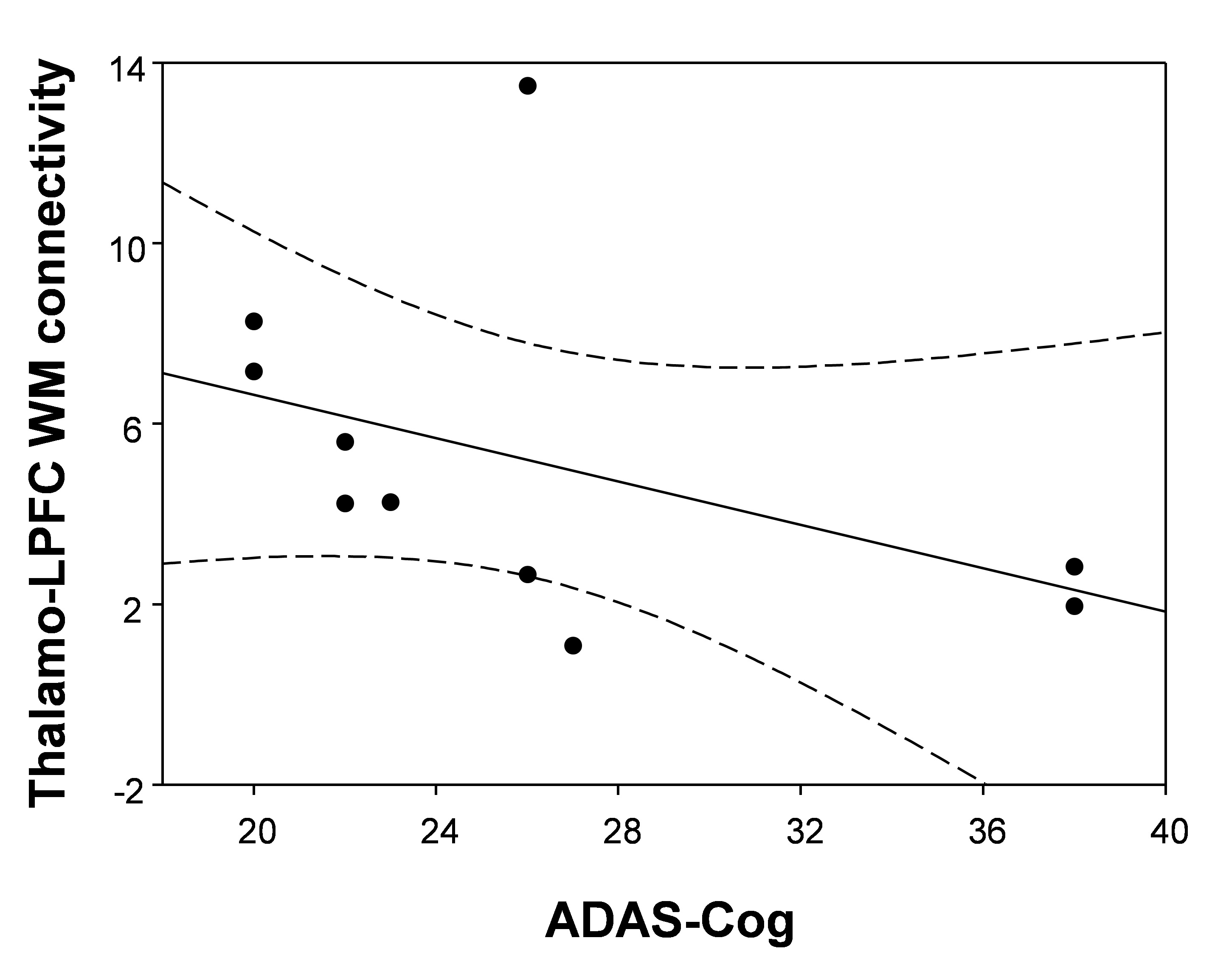
The donepezil-treated patients showed the increased thalamo-medial temporal cortex (MTC) white matter connectivity after 6 months of donepezil treatment (Fig. 2). Compared with untreated patients with AD, donepezil-treated patients with AD showed significantly higher GM volumes in the inferior frontal gyrus (IFG) and inferior temporal gyrus (ITG) (Fig. 3). The GM volumes of the IFG (Spearman’s rho = 0.67, p = 0.035) and ITG (Spearman’s rho = 0.73, p = 0.017) in treated patients were positively correlated with MMSE scores (Fig. 4). The thalamo-LPFC WM connectivity in treated patients was negatively correlated with ADAS-cog scores (Spearman’s rho = -0.68, p = 0.030) (Fig. 5). Our findings suggest that the increased thalamo-MTC white matter connectivity and enhanced GM volumes of the IFG and ITG in donepezil-treated patients can be attributed to the treatment.
Conclusion
This study has demonstrated the altered thalamo-cortical white matter connectivity and GM volume after donepezil treatment in patients with AD. These findings will be useful for constraining theories on the etiology of AD and the mode of action of anticholinesterases in treating it.Acknowledgements
This research was supported by National Research Foundation of Korea grants funded by the Korea government (Ministry of Science and Information and Communication Technology) (2018R1A2B2006260 and 2018R1C1B6005456), and Chonnam National University (CNU) Research Fund for CNU distinguished research emeritus professor (2017-2022).References
1. Hashimoto M, Kazui H, Matsumoto K, et al. Does donepezil treatment slow the progression of hippocampal atrophy in patients with Alzheimer's disease? Am J Psychiatry. 2005;162:676-682.
2. Birks J. Cholinesterase inhibitors for Alzheimer's disease. Cochrane Database Syst Rev. 2006;1:CD005593.
3. Modrego PJ, Fayed N, Errea JM, et al. Memantine versus donepezil in mild to moderate Alzheimer's disease: a randomized trial with magnetic resonance spectroscopy. Eur J Neurol. 2010;17:405-412.
4. Kasa P, Papp H, Kasa P, Jr., et al. Donepezil dose-dependently inhibits acetylcholinesterase activity in various areas and in the presynaptic cholinergic and the postsynaptic cholinoceptive enzyme-positive structures in the human and rat brain. Neuroscience. 2000;101:89-100.
5. Scali C, Casamenti F, Bellucci A, et al. Effect of subchronic administration of metrifonate, rivastigmine and donepezil on brain acetylcholine in aged F344 rats. J Neural Transm. 2002;109:1067-1080.
6. Ikuta T, Shafritz KM, Bregman J, et al. Abnormal cingulum bundle development in autism: a probabilistic tractography study. Psychiatry Res. 2014;30:63-68.
7. Cho KI, Shenton ME, Kubicki M, et al. Altered Thalamo-Cortical White Matter Connectivity: Probabilistic Tractography Study in Clinical-High Risk for Psychosis and First-Episode Psychosis. Schizophr Bull. 2016;42:723-731.
Figures
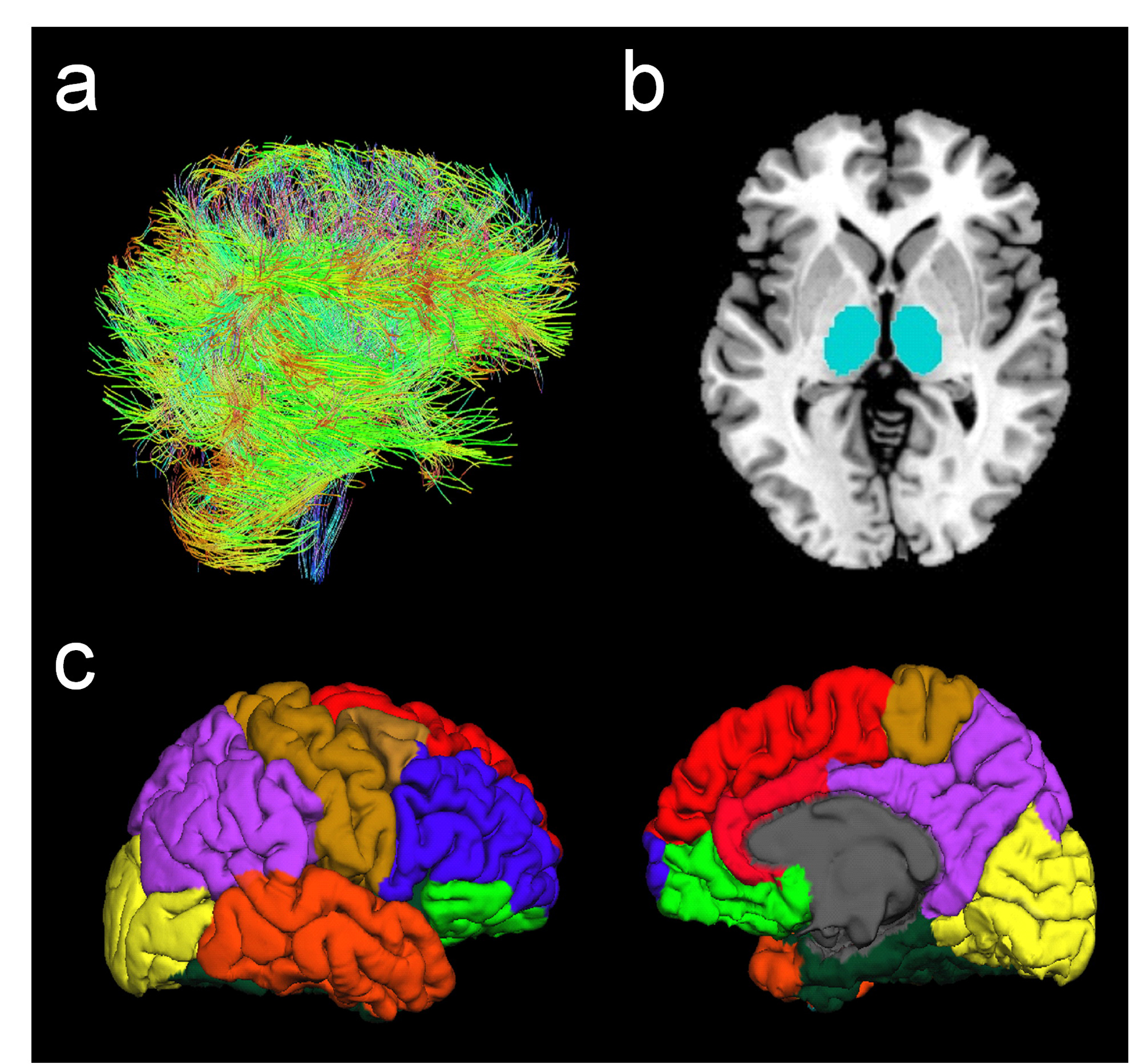
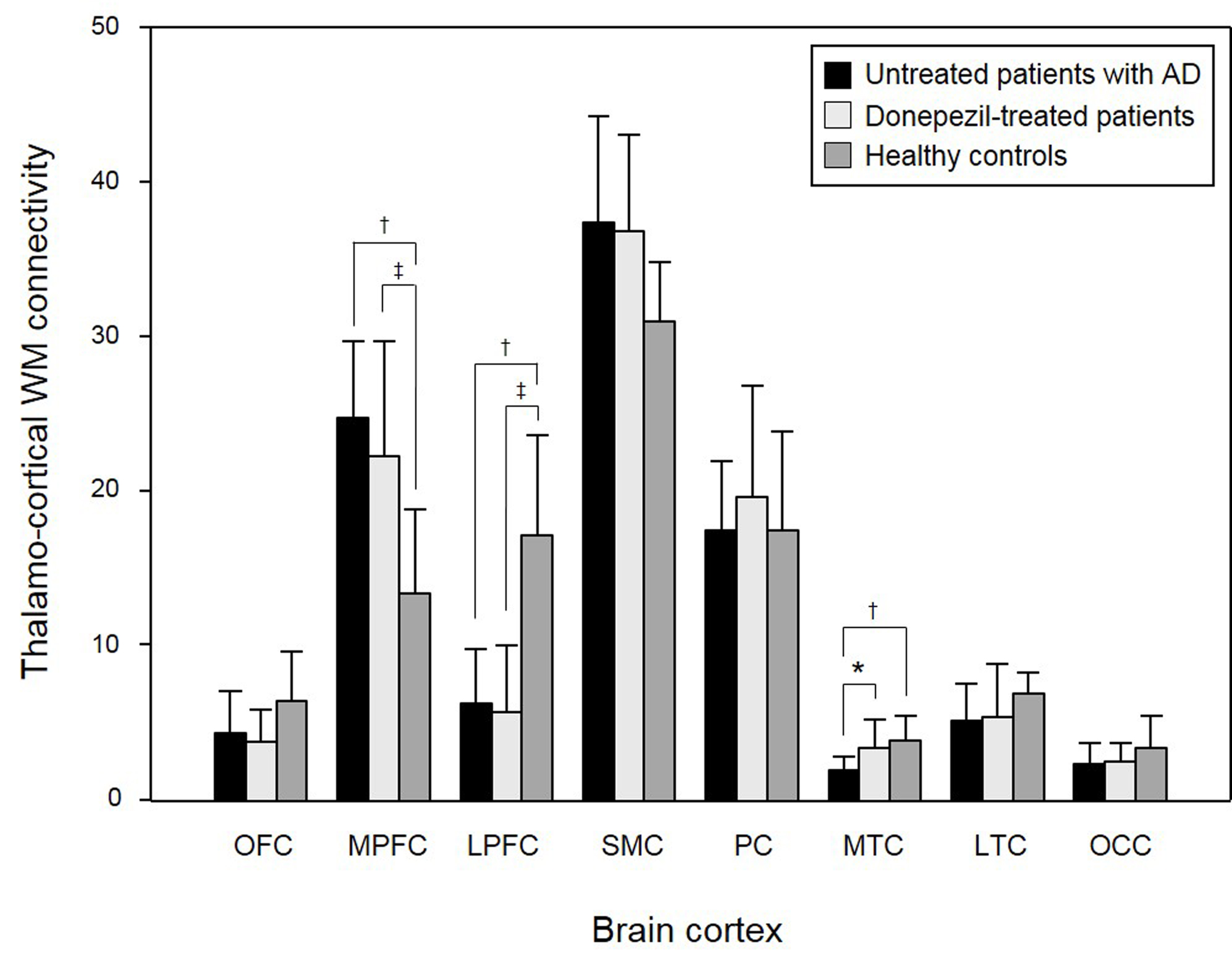
Fig. 2. Comparison of the mean white matter connectivities between thalamus and each cortex in untreated patients with AD, donepezil-treated patients with AD, and healthy controls. Orbitofrontal cortex: OFC, medial prefrontal cortex: MPFC, lateral prefrontal cortex: LPFC, sensorimotor cortex: SMC, parietal cortex: PC, medial temporal cortex: MTC, lateral temporal cortex: LTC, occipital cortex: OC.
†significant difference (Mann-Whitney U; p < 0.05) between untreated patients and healthy controls.
‡significant difference (Mann-Whitney U; p < 0.05) between donepezil-treated patients and healthy controls.
*significant difference (Wilcoxon's signed-ranks; p < 0.05) between untreated patients and donepezil-treated patients.