3043
Sex and Hemispheric Dependent DTI-based Network Analysis of an Alzheimer’s Disease Mouse Model1National High Magnetic Field Laboratory, Florida State University, Tallahassee, FL, United States, 2Chemical & Biomedical Engineering, FAMU-FSU College of Engineering, Tallahassee, FL, United States, 3Biomedical Engineering, Johns Hopkins University, Baltimore, MD, United States, 4Applied Mathematics & Statistics, Johns Hopkins University, Baltimore, MD, United States, 5Wallace H. Cooulter Department of Biomedical Engineering, Georgia Institute of Technology, Atlanta, GA, United States, 6Wallace H. Coulter Department of Biomedical Engineeing, Emory University, Atlanta, GA, United States
Synopsis
This study utilizes DTI and graph theory as a novel way for early detection of pathology and connectivity changes related to Alzheimer’s Disease. As a function of phenotype, age and sex, DTI studies were performed on APP/PS1 mouse brains and age-matched wild type controls at 11.75 T. Current hemisphere-dependentdata shows differences between hemispheres within age and phenotype for the parameters observed.
Introduction
Alzheimer’s Disease (AD) is the most common form of dementia, characterized by memory loss and changes in behavior1. The most prevalent preclinical model of AD is the APP/PS1 mouse expressing human genes for amyloid precursor protein (APP) and presenilin-1 (PS1). Clinically, MRI is used to diagnose AD by means of volumetrics, mainly focusing on hippocampal atrophy2. In this study, diffusion tensor imaging (DTI) is applied to the 5xFAD variant of the APP/PS1 mouse model. At multiple early time points, network theory was employed to examine alterations in structural connectivity as a function of age, sex and across hemispheres compared to wild-type controls. The investigation of hemispheric dependence in the loss of neural connectivity was partially motivated by a previous study that showed possible hemispheric differences in vascular pathology related to AD3.Methods
Image datasets were acquired using preserved mouse brains (4% paraformaldehyde) from male and female specimens either expressing the APP/PS1 phenotype or an age-matched wild type. Brains were harvested at 1, 2, 4 and 6 months (N=5 per age, gender and phenotype). Using an 11.75-T magnet, DTI data was acquired with a multi-slice 2D spin echo sequence using 18 diffusion encoding directions (nominal b = 400 s/mm2) and four unweighted acquisitions for which Δ=21 ms and δ=3 ms. Two to three brains were imaged simultaneously with an in-plane resolution of 100x100 mm, matrix size of 256x256, slice thickness of 500mm, repetition time of 2 s and echo time of 30 ms. With 15 averages and a 47-hr total acquisition time, high signal-to-noise ratios of 60:1 were obtained. DTI data were analyzed using DSI Studio4 to generate tracts using the following parameters: Threshold=0.10, Angular Threshold=60o, Step Size=0.05mm, Minimum Length=1mm, Maximum Length=25mm, and termination after 106 seeds. Adjacency matrices were created by running whole brain tracking and placing regions of interest (ROIs) within developed tracts and generating a pass through matrix. Network matrices then were utilized by GEPHI to obtain network properties. A hemisphere-dependent analysis was completed to indicate any changes that may exist. A one-way ANOVA with a least significant difference post-hoc test (p<0.05) was used to determine statistical significance among samples.Results & Discussion
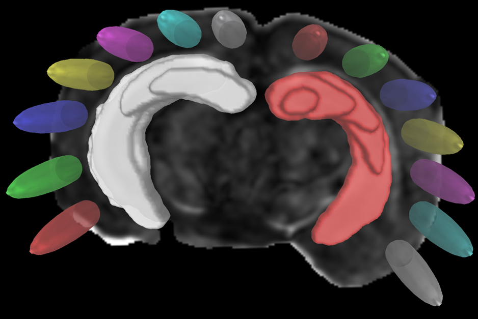
Four
main areas of focus were the Piriform Area, Temporal Lobe, Parietal Lobe and
Hippocampus. These regions divided into
left and right hemispheres increased the number of regions to eight. Structural
connectivity work was assessed using 14 equally spaced prolate spheroidal ROIs
that were positioned in cortical regions, as well as manually segmented nodes
representing the left and right hippocampus, to quantify connectivity both
locally and globally [Figure 1]. Tracts
were measured between each individual node in order to derive quantifiable
adjacency matrices that correspond to neural graphs. The analysis was done
using a weighted approach to study the structural connectivity changes in the
models.
Impacts
of aging can be seen in closeness and harmonic centrality when hemispheric
differences are not considered. Weighted degree and clustering in older samples
show impacts of phenotype. In younger samples, impacts of phenotype can be seen
in eccentricity as well as closeness and harmonic centrality.
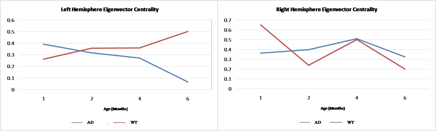
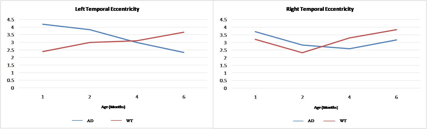
When
comparing left and right hemisphere differences for certain network metrics, a
divergent pattern with age for female transgenic (decreases) versus wild-type(increases)
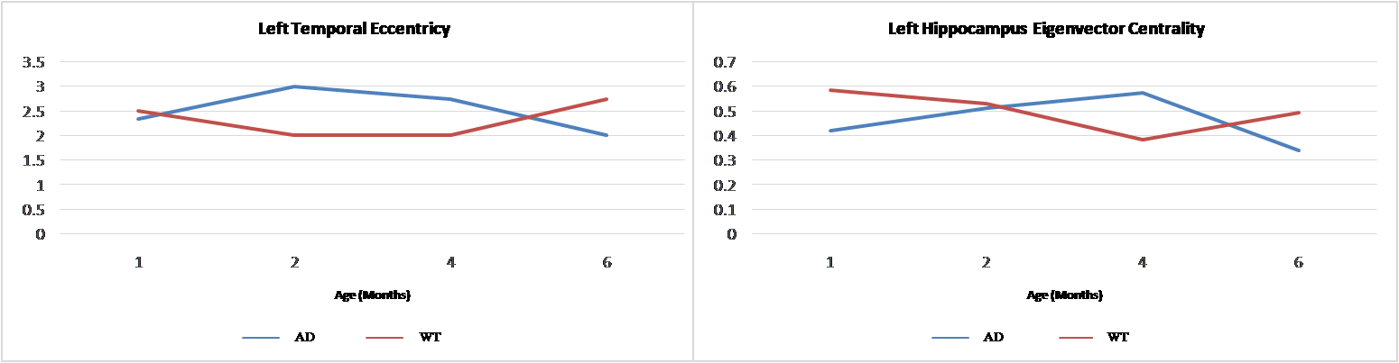
developed, localized to the left hemisphere [Figures 2,3]. Male specimens generally did not show this hemispheric
trend, instead wild-type network metrics either remained stable or decreased
until 4 months before rebounding while transgenic metrics increased until 4 months
then decreased [Figure 4].Conclusion
This research utilizes network theory and MRI to detect and classify the progression of Alzheimer’s Disease, potentially providing early hallmarks of structural connectivity changes that may be impacted by sex. Such classifications may assist in defining treatment regiments earlier in disease progression, possibly before hallmark symptoms present. Additionally, this work will help to expand the application of DTI and network theory to identification and assessment of progression of other neurodegenerative diseases.Acknowledgements
This work was funded by NSF (DMR-1157490 & DMR-1644779), the State of Florida, the National High Magnetic Field Laboratory User Collaborations Grant Program, and NIH (R01 NS102395).
References
1. U.S Department of Health & Human Services.www.alzheimers.gov.
2. Tang, X., Qin, Y., Wu, J., Zhang, M., Zhu, W., & Miller, M. I. 2016. Shape and diffusion tensor imaging based integrative analysis of the hippocampus and the amygdala in Alzheimer's disease. Magnetic resonance imaging. 34(8):1087-1099.
3. Giannakopoulos, P., Kövari, E., Herrmann, F. R., Hof, P. R., & Bouras, C. 2008. Interhemispheric distribution of Alzheimer disease and vascular pathology in brain aging. Stroke, 40(3), 983-6.
4. Yeh F, Verstynen TD, Wang Y, Fernández-Miranda JC, Tseng WI. 2013. Deterministic diffusion fiber tracking improved by quantitative anisotropy. PLoS One. 8(11): e80713.
Figures