3030
Characterization of Diffusion Anisotropy Alterations Associated with Dilated Virchow Robin Spaces in Simple Febrile Seizure Children between 12 and 48 MonthsMustafa Salimeen Abdelkareem1, Xianjun Li1, Miaomiao Wang1, Congcong Liu1, Habib Tafawa1, Anuja Pradhan1, Martha Singh1, Xiaocheng Wei2, Guanyu Yang3, and Jian Yang1,4
1Department of Radiology, The First Affiliated Hospital of Xi’an Jiaotong University, Xi’an, China, 2MR Research China, GE Healthcare, Beijing, China, Xi’an, China, 3Xi'an AccuRad Network and Technology Co. Ltd., Xi'an, China, Xi’an, China, 4Department of Biomedical Engineering, School of Life Science and Technology, Xi’an, China
Synopsis
Dilated Virchow Robin spaces (dVRs) are common in febrile seizure (FS) patients. However, little is known about how dVRs affect the white matter structure in developing brains. This study aimed to characterize the anisotropy alterations in white matter associated with dVRs in simple FS children by using fractional anisotropy (FA). Through inter-group comparisons, FA was larger in simple FS with dVRs children than that in FS without dVRs and control groups. Significant positive correlations between FA and VRs count, seizure duration were found. These results suggest that dVRs can affect the structure of white matter by increasing FA values.
INTRODUCTION
Febrile seizures (FS) are common childhood seizures and occur in associated with a high body temperature its peak age is 18 months. Simple FS is seizure duration lasting less than 15 minutes1. Virchow-Robin spaces (VRs) are surrounding perforating cerebral vessels can be seen on magnetic resonance imaging (MRI) and its isointense to cerebrospinal fluid (CSF) in MRI sequences2. Dilated Virchow robin spaces (dVRs) were found in seizures children3. Till to now, the effect of dVRs on white matter structure is not well understood. Recently there are evidences revealing that dVRs are associated with pathological conditions4. The dVRs are considered as mass effect in alteration of FA value5. However, few studies address the effect of dVRs on FA value alterations, such as simple FS children. Therefore, this study aimed to evaluate the effect of dVRs on FA value changes in simple FS children through diffusion tensor imaging (DTI) and tract-based spatial statistics (TBSS).MATERIALS AND METHODS:
The study was approved by the local institutional review board. Written informed consents were obtained from the parents. Fifty simple FS with dVRs, 20 simple FS without dVRs, and 35 control children were evaluated retrospectively. The children with simple FS were diagnosed by two pediatric neurologists. MRI examinations were performed using a 3T scanner (Signa HDxt, GE Healthcare, Milwaukee, Wisconsin, USA) with an 8-channel head coil. We acquired T2 weighted sequence (TR/TE, 4200ms/120 ms; matrix, 256*256; slice thickness, 2.5 mm; FOV, 240 mm) and T2 FLAIR sequence (TR/TE, 8600 ms/165 ms; matrix, 288*224; slice thickness, 5 mm; FOV, 240 mm). DTI parameters were as follows: 30 gradient directions; b values = 0 and 600 s/mm2; TR/TE = 11000/69.5ms; slice thickness = 2.5 mm without spaces; FOV = 240 mm; and matrix size = 128 × 128. FMRIB software library (FSL, www.fmrib.ox.au.uk/fsl) was used for processing DTI data. FA maps were obtained after the brain extraction and the eddy current correction. Linear and non-linear image registrations were utilized for alignment of the FA maps of all subjects to a selected FA map. An averaged image of the co-registered FA maps was created as the target map. Then, FA maps of all subjects were registered to the target map. The mean FA map and mean FA skeleton were created. The aligned FA map of each subject were projected onto the mean FA skeleton (threshold = 0.15). Voxel-wise statistical analysis was performed to assess the inter-group differences in FA with the threshold-free cluster enhancement and family-wise error correction. Regional values were extracted from the areas with significant differences revealed by TBSS. Categorical variables were analyzed by using Chi-square test in the SPSS software (Version 21.0; IBM, Armonk, New York, USA). The continuous variables were analyzed by using Mann-Whitney U test. P values less than 0.05 were considered statistically significant.RESULTS
There was significant difference between simple FS with dVRs and simple FS without dVRs in seizure duration (Table 1), (P <0.001) and white blood count (WBCs), neutrophil (P=0.007), (P = 0.008) respectively (Table1). Compared with simple FS without dVRs group, no significant difference in the time between controlling the seizure and MRI scanning was found in simple FS with dVRs group (P= 0.741) (Table1). TBSS analysis demonstrates significant increased FA values for the simple FS patients with dVRs, compared with simple FS without dVRs and control groups (Figure 1 and 2). Positive significant correlations were found in terms of FA and seizure duration(P<0.0001) and VRs count (P=0.0001) (Figure 2).DISCUSSION
For simple FS patients with dVRs, FA value increased in the brain white matter (WM). Such changes of FA value may indicate maturation of WM microstructure, detailed as increased axon density, axonal diameter, directionality myelination and decreased water content6. Therefore, above changes may lead to the changes of DTI metrics in the majority of WM areas. Dilated VRs act as mass effect, compress water molecules in WM, and improve it is rearrangement order, and accelerate WM development7 extracellular fluid accumulation, decrease secondary to this mass effect, suggesting augmented diffusion anisotropy. This suggests high density of normal axons, as corroborated by increased index of the fiber density8. The contribution of this work is to provide information that frequent dVRs may be lead to FA changes in simple FS preschool children.CONCLUSION
This study provides an evidence that dVRs can affect the structure of WM by increasing FA values in simple FS children.Acknowledgements
The study was supported by the National Key Research and Development Program of China (2016YFC0100300), National Natural Science Foundation of China (No.81171317, 81471631, 81771810), the 2011 New Century Excellent Talent Support Plan of the Ministry of Education, China (NCET-11-0438), the Clinical Research Award of the First Affiliated Hospital of Xi’an Jiao Tong University (No. XJTU1AF-CRF-2015-004) , the Fundamental Research Funds for the Central Universities (xjj2018265), the Fundamental Research Funds of the First Affiliated Hospital of Xi'an Jiaotong University (2017QN-09).References
1. Oh JS CJ, Jeong H. Interleukin-1b interleukin-1 receptor antagonist levels in children with febrile seizures. J Korean Child Neurol Soc 2016; 24:84–8. 2. Rebecca Emily Feldman, John Watson Rutland, Madeline Cara Fields, Lara Vanessa Marcuse, Puneet S. Pawha, Bradley Neil Delman, Priti Balchandani, Quantification of perivascular spaces at 7 T: A potential MRI biomarker for epilepsy; R.E. Feldman et al. / Seizure 54 (2018) 11–18 3. Wuerfel J, Haertle M, Waiczies H, et al. Perivascular spaces: MRI marker of inflammatory activity in the brain? Brain 2008; 131:2332–2340. 4.GroeschelS, BrockmannK, HanefeldFVirchowRobin spaceson magnetic resonance images of children with adrenoleukodystrophy, Eur J Paediatr Neurol. 2007 May;11(3):142-5. 5. Alberto Cacciola, Rocco Salvatore Calabrò, Antonio Costa, Antonino Naro,Demetrio Milardi, Daniele Bruschetta:Enlarged Virchow-Robin spaces in a young man:a constrained spherical deconvolution tractography study;Acta Biomed 2017; Vol. 88, N. 3: 319-324 6. Dubois J, et al. Assessment of the early organization and maturation of infants’ cerebral white matter fiber bundles: a feasibility study using quantitative diffusion tensor imaging and tractography. Neuroimage, 2006, 30(4): 1121-1132 7. Feldman, H.M., Lee, E.S., Yeatman, J.D., Yeom, K.W., 2012. Language and reading skills in school-aged children and adolescents born preterm are associated with white matter properties on diffusion tensor imaging. Neuropsychologia 50, 3348–3362. https://doi.org/10.1016/j.neuropsychologia.2012.10.014. 8. Stadlbauer A, Nimsky C, Gruber S, et al. Changes in fiber integrity, diffusivity, and metabolism of the pyramidal tract adjacent to gliomas: a quantitative diffusion tensor fiber tracking and MR spectroscopic imaging study. AJNR Am J Neuroradiol 2007; 28:462-469.Figures
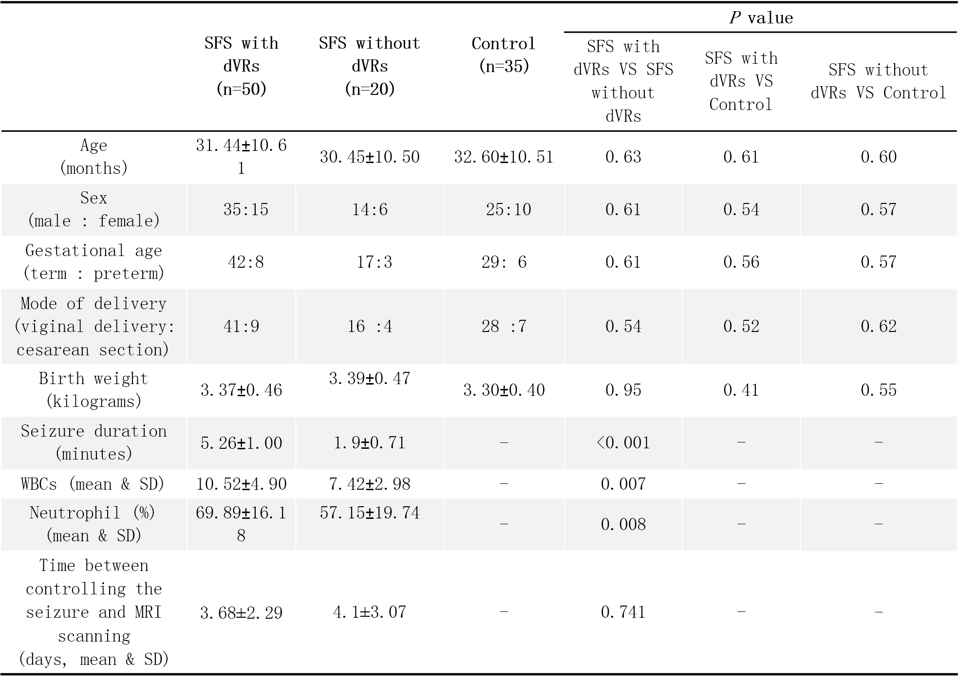
Patient clinical and demographic data
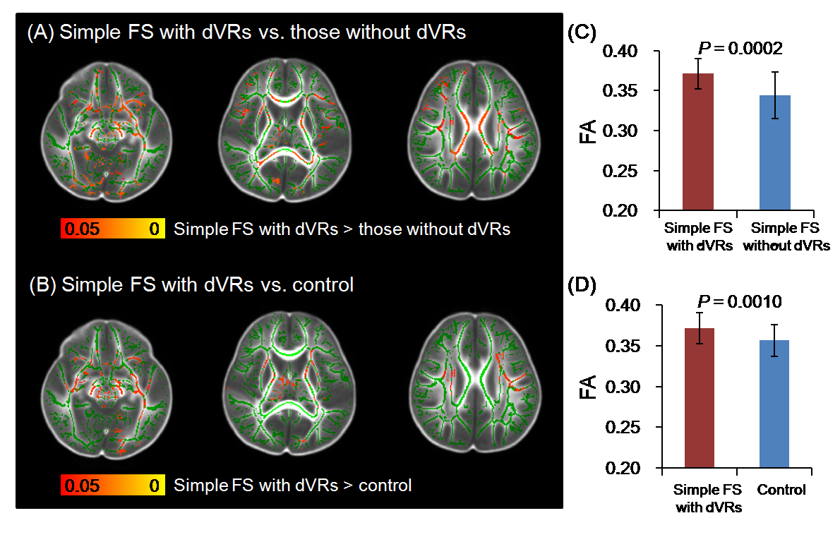
Inter-group comparisons by using TBSS and ROI
analyses
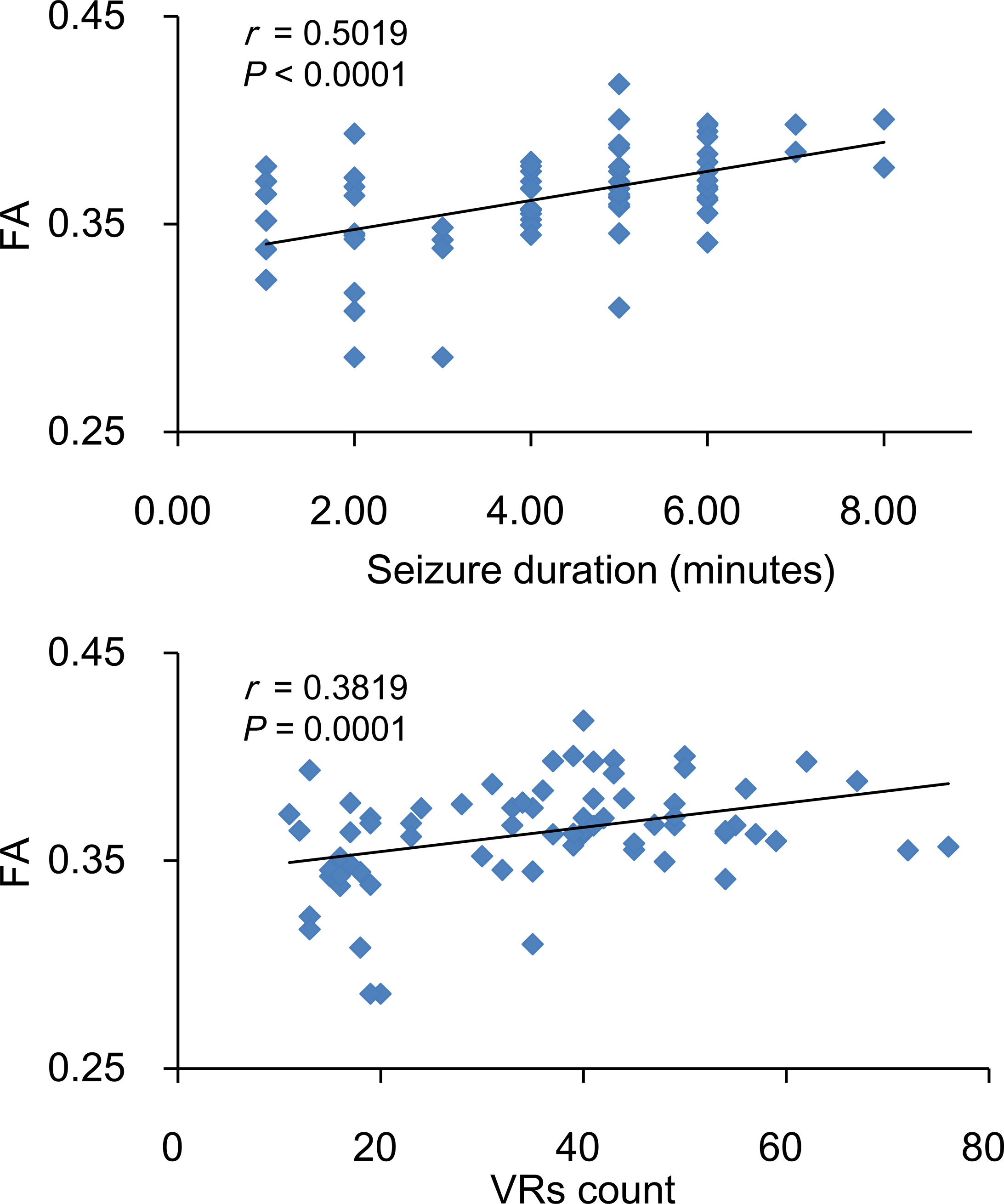
Correlation between FA and seizure duration (top) and VRs count.
(bottom).