3029
What’s shape got to do with it? Exploring subcortical shape and volume alterations in youth with congenital heart disease1Advances in Brain and Child Development Research Laboratory, Research Institute of the McGill University Health Centre, Montreal, QC, Canada, 2Integrated Program in Neuroscience, McGill University, Montreal, QC, Canada, 3Department of Cardiology, Montreal Children’s Hospital, McGill University Health Centre, Montreal, QC, Canada, 4Department of Medical Imaging, Montreal Children’s Hospital, McGill University Health Centre, Montreal, QC, Canada, 5MR Clinical Science, Philips Healthcare, Markham, ON, Canada, 6Department of Pediatrics, Montreal Children’s Hospital, McGill University Health Centre, Montreal, QC, Canada, 7Computational Brain Anatomy Laboratory, Cerebral Imaging Centre – Douglas Mental Health University Institute, Verdun, QC, Canada
Synopsis
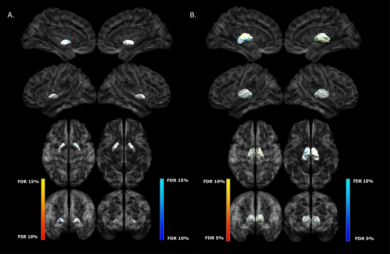
Congenital heart disease (CHD) is a leading cause of long-lasting neurodevelopmental impairment. Evaluating subtle neuroanatomical variation using magnetic resonance imaging data has been shown to be sensitive for capturing morphometric signatures related to neurodevelopmental disorders. In this study, we found morphometric differences in subcortical structures of youth with CHD even in the absence of volumetric differences. While we did not find any significant morphometric differences between groups for the striatum, we did find smaller surface area and inward bilateral inward displacement across the lateral surfaces of the globus pallidus and the thalamus in the CHD group compared to controls.
Introduction
Advanced structural magnetic resonance imaging (MRI) is increasingly used to provide information about the shape of brain structures of interest. Shape analysis has the potential to further identify brain differences that cannot be captured by volumes alone, such as bending, flattening or focal surface area changes. Morphometry has been shown to provide a sensitive marker for subtle structural brain differences in various neurodevelopmental disorders, such as autism spectrum disorder, attention deficit and hyperactivity disorder and child onset schizophrenia1-3. Congenital heart disease (CHD) is a leading cause of long lasting neurodevelopmental impairment4. Brain injury and neurodevelopmental delay is well documented in children with CHD, however, there are a limited number of studies which have evaluated brain structure integrity in adolescents born with CHD. It has been reported that even in the absence of brain anomalies on conventional MRI, adolescents with CHD presented with worse neurocognitive function than healthy peers5. While conventional MRI provides insight into the nature and frequency of evident brain abnormalities, it does not capture the subtle structural brain differences that could be underlying many of the neurodevelopment deficits observed in this population. Impairments in executive function and motor skills are among the most prevalent in individuals with CHD6. Many subcortical structures such as the globus pallidus, striatum and thalamus play crucial roles in higher-order cognitive processing and motor functions. Since comprehensive evaluation of the subcortical structures is lacking in the CHD population, the objective of this project was to compare morphometry and volume of the globus pallidus, striatum, and thalamus between youth born with CHD and healthy peers.Methods
We recruited youth between 16-24 years of age who were born with CHD having undergone cardiopulmonary bypass surgery before two years of age and healthy controls of the same age. All participants underwent a brain MRI on a Philips 3T MR System using a 32-channel head coil to acquire 1 mm isotropic 3D T-1 weighted images (TR/TE/flip angle = 8.1ms/3.7ms/8º). All raw images were preprocessed using the minc-bpipe-library7,8. This pipeline corrects for bias field contrast inhomogeneity using the N4ITK algorithm9 and performs a brain extraction based on nonlocal segmentation technique (BEaST) [10]. Total brain volume (TBV) estimates were acquired from the BEaST mask 10. To delineate the subcortical structures, all pre-processed images were post-processed using The Multiple Automatically Generated Templates for different algorithm (https://github.com/CobraLab/MAGeTbrain)11. Images were submitted to the morphometric branch of MAGeT-Brain to yield vertex-wise surface area maps for each segmentation. Descriptive statistics were used to characterize the sample. Subjects’ characteristics were compared between groups using independent t-tests for continuous variables and chi-square tests for categorical variables. Group differences in shape and volume were calculated with general linear modelling using TBV as a covariate. False discovery rate was used to correct for multiple comparisons.Results
In this ongoing cross-sectional study, brain MRIs have been collected in 49 youth born with CHD and 48 healthy controls (mean age of 20.3 years). There were no significant differences with respect to age, sex or BMI between the two groups. Smaller surface area and inward bilateral inward displacement across the lateral surfaces of the globus pallidus was concentrated anteriorly in the CHD group compared to controls (q<0.15). On the lateral surfaces of the bilateral thalami, we found regions of both increased and decreased surface area, as well as, inward and outward displacement in the CHD group compared to controls (q<0.15) (see figure 1). We did not find any significant morphometric differences between groups for the striatum. We found that only the globus pallidus showed a significant volume reduction (q<0.01) in the CHD group when compared to controls, suggesting that volumetric differences were not underlying the morphometric differences observed.Discussion
Previous studies conducted in youth with CHD have explored the volumetric properties of subcortical structures exclusively. Here we report differences in morphometric measurements for the first time. In line with studies which have looked at subcortical morphometry in other populations with neurodevelopmental impairments, our findings suggest that volume alone may not be sufficient to detect subtle structural brain differences. Future studies will focus on structure-function relationships to better understand how the morphometric differences in subcortical structures relate functional outcomes of survivors of CHD.Conclusion
This study provides the first comprehensive evaluation of the subcortical structures of youth with CHD. The morphometric differences identified here may expand our understanding of the underlying mechanisms of maldevelopment and neurodevelopmental outcomes observed in individuals with CHD.Acknowledgements
The study was supported by the start-up funds from the Research Institute of the McGill University Health Centre and McGill University.
At the time of the study, K.F. received financial support from McGill University’s Faculty of Medicine Internal Studentship Award, given as: Joseph Schubert Memorial Award & Jeannette and Abram Victor Fellowship Award and the Research Institute of the McGill University Health Centre ― Desjardins Studentship in Child Health Research.
We would also like to thank the youth and their families for their participation, and the clinicians, technologists and research assistants for their implication in this study.
Computations were performed on the Niagara supercomputer at the SciNet HPC Consortium. SciNet is funded by: the Canada Foundation for Innovation under the auspices of Compute Canada; the Government of Ontario; Ontario Research Fund - Research Excellence; and the University of Toronto.
References
1. Chakravarty, M.M., et al., Striatal shape abnormalities as novel neurodevelopmental endophenotypes in schizophrenia: a longitudinal study. Hum Brain Mapp, 2015. 36(4): p. 1458-69.
2. Schuetze, M., et al., Morphological Alterations in the Thalamus, Striatum, and Pallidum in Autism Spectrum Disorder. Neuropsychopharmacology, 2016. 41(11): p. 2627-37.
3. Shaw, P., et al., Mapping the development of the basal ganglia in children with attention-deficit/hyperactivity disorder. J Am Acad Child Adolesc Psychiatry, 2014. 53(7): p. 780-9.e11.
4. Marino, B.S., et al., Neurodevelopmental outcomes in children with congenital heart disease: evaluation and management: a scientific statement from the American Heart Association. Circulation, 2012. 126(9): p. 1143-72.
5. Bolduc, M.E., et al., Structural brain abnormalities in adolescents and young adults with congenital heart defect: a systematic review. Dev Med Child Neurol, 2018. 60(12): p. 1209-1224.
6. Marelli, A., et al., Brain in Congenital Heart Disease Across the Lifespan: The Cumulative Burden of Injury. Circulation, 2016. 133(20): p. 1951-62.
7. Sadedin, S.P., B. Pope, and A. Oshlack, Bpipe: a tool for running and managing bioinformatics pipelines. Bioinformatics, 2012. 28(11): p. 1525-6.
8. Vincent, R.D., et al., MINC 2.0: A Flexible Format for Multi-Modal Images. Front Neuroinform, 2016. 10: p. 35.
9. Tustison, N.J., et al., N4ITK: improved N3 bias correction. IEEE Trans Med Imaging, 2010. 29(6): p. 1310-20.
10. Eskildsen, S.F., et al., BEaST: Brain extraction based on nonlocal segmentation technique. NeuroImage, 2012. 59(3): p. 2362-2373.
11. Chakravarty, M.M., et al., Performing label-fusion-based segmentation using multiple automatically generated templates. Human Brain Mapping, 2013. 34(10): p. 2635-2654.
Figures