3027
Altered global white matter microstructure and structural brain connectivity in children born with extremely low birth weight1HUS Medical Imaging Center, Radiology, University of Helsinki and Helsinki University Hospital, Helsinki, Finland, 2Department of Neuroscience and Biomedical Engineering, Aalto University, Espoo, Finland, 3Turku Brain and Mind Center, University of Turku, Turku, Finland, 4Children´s Hospital, Helsinki University Hospital, Helsinki, Finland, 5Pediatrics, Department of Clinical Sciences, Lund University, Lund, Sweden
Synopsis
Using diffusion MRI, we investigated white matter microstructure and structural brain connectivity in 11-year old children born with extremely low birth weight (ELBW) in comparison with full-term born children. Microstructural white matter properties were investigated within the tract skeleton, and constrained spherical deconvolution based probabilistic tractography and gray matter parcellation were used to reconstruct structural brain connectivity networks. We found decreased integrity and complexity of the white matter microstructure in ELBW, and increased segregation of the structural brain connectivity networks. In addition, the microstructural changes were associated with the administration of antenatal corticosteroids and with retinopathy of prematurity.
Introduction
Preterm birth rate in Europe varies between 5-10% of all births1. As the brain rapidly grows and develops during the last trimester of gestation2, very preterm infants are at greater risk for abnormalities in brain development3 leading to motor, cognitive, and social deficits such as cerebral palsy, learning disabilities, and autism spectrum disorder4-7. Therefore, it is important to understand the neurobiological abnormalities related to prematurity, and to recognize the neonatal risk factors leading to neurodevelopmental impairments.
Diffusion-weighted (DW) magnetic resonance imaging (MRI)8 has enabled noninvasive investigation of the neural tracts and their microstructural properties in vivo. In this study, we investigated white matter microstructure and structural brain connectivity networks in 11-year old children born with extremely low birth weight (ELBW) from Helsinki region, a geographically defined subgroup of a national ELBW cohort9, and compared them to full-term (FT) born children.
Methods
Diffusion MRI data were acquired from 37 ELBW children (aged 11.3±1.2 years, 25 born extremely preterm <28 gestational weeks) and 29 FT children (aged 11.6±0.7 years) with a Philips Achieva 1.5T machine by using 20 gradient orientations and a b‑value of 800 s/mm2 with a resolution of 1.75 mm × 1.75 mm × 2 mm. T1‑weighted MRI data were acquired with a resolution of 0.94 mm × 0.94 mm × 1 mm. The study protocol was approved by the Ethics Committee of Helsinki and Uusimaa Hospital District, Finland. The parents and the child gave written informed consent before the investigation.
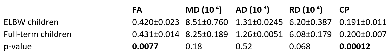
The DW data were corrected for subject motion10, eddy current induced11, and echo planar imaging distortions12. White matter tract skeleton was reconstructed as introduced in tract-based spatial statistics (TBSS)13. Global microstructural changes in fractional anisotropy (FA), mean diffusivity (MD), axial diffusivity (AD), radial diffusivity (RD), and coefficient of planarity (CP) were investigated14.
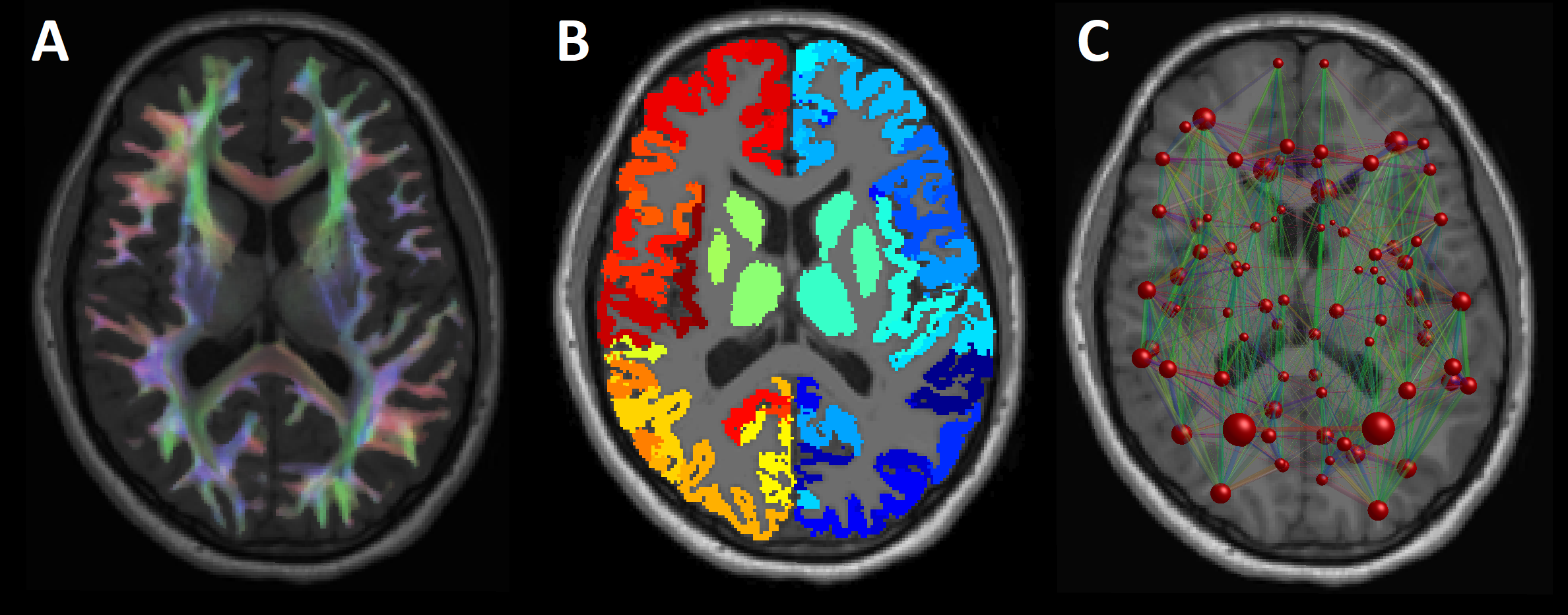
We investigated the structural brain connectivity networks by using graph theoretical analysis15. First, we estimated the fiber orientation distributions with constrained spherical deconvolution (CSD)16 by using up to fourth order spherical harmonics and performed probabilistic streamlines tractography in MRtrix17-18. Cortical parcellation of the T1-weighted images was performed in FreeSurfer19 by using the Desikan-Killiany atlas20, combined with the subcortical gray matter structures segmented with FIRST21 in FSL22. Then, the two endpoints of each streamline were assigned to the gray matter areas, resulting in structural connectivity networks, presented in Figure 1, in which the nodes represent the gray matter areas and the edges represent the number of streamlines between the nodes.
We compared global microstructural and network properties in ELBW children to FT control subjects and looked for associations between the properties with the following ante- and neonatal factors: gestational age, birthweight, antenatal corticosteroid administration, twin pregnancy, intrauterine growth restriction, vaginal delivery, respiratory distress syndrome, neonatal sepsis, retinopathy of prematurity stage III or higher, and need of oxygen at the age corresponding to 36 gestational weeks.
Results
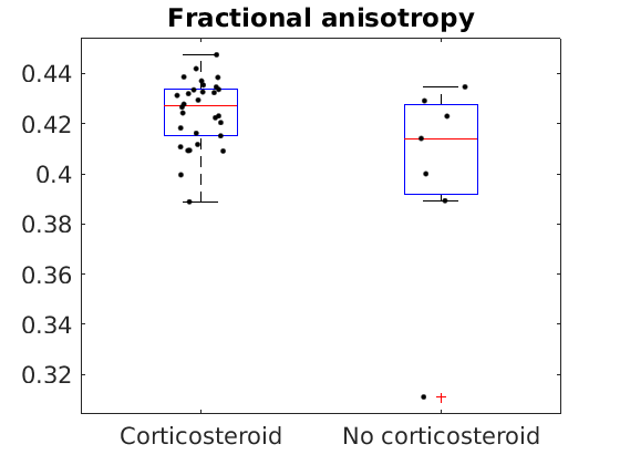
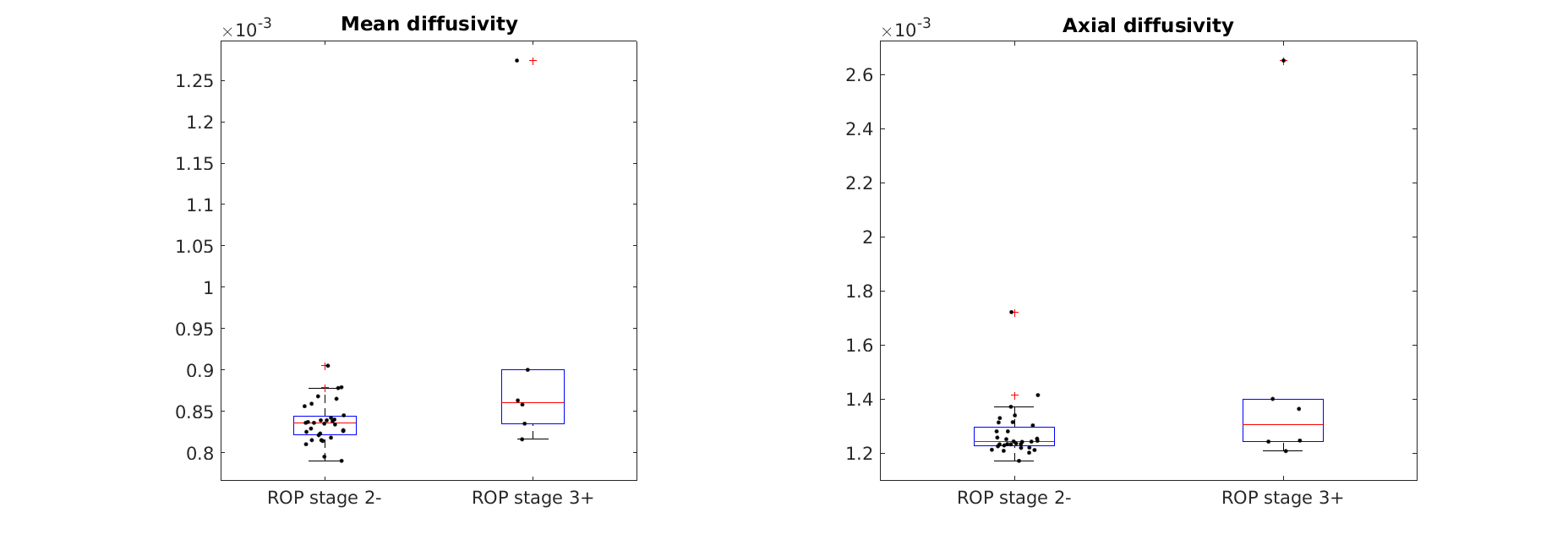
We found significantly decreased FA and CP in ELBW children compared to FT children, as shown in Table 1. Furthermore, the ELBW children whose mothers did not receive antenatal corticosteroid had decreased FA compared to those treated, as shown in Figure 2. In addition, the children with retinopathy of prematurity stage III or higher had increased MD and AD compared to those with no or lesser retinopathy degree, as shown in Figure 3. However, there were no significant correlations between gestational age or birth weight and the microstructural measures in ELBW children. No associations were found with the other investigated factors.
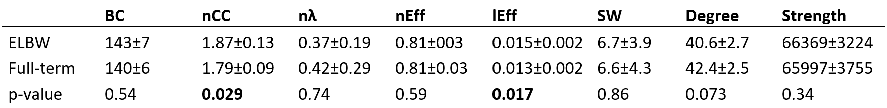
We found increased normalized clustering coefficient and local efficiency in ELBW children compared to FT children, as shown in Table 2. No correlations with the gestational age or birth weight were found, but vaginal delivery was associated with increased local efficiency (p=0.035).
Discussion
We found global microstructural and connectivity abnormalities in the preterm group compared to FT children. A decrease in FA in ELBW children suggests decreased white matter integrity or myelination23, while a decrease in CP suggests a decrease in the complexity of the white matter organization14. Increased clustering coefficient and local efficiency were found in ELBW children, which indicates increased segregation of the structural brain connectivity networks15. In addition, the microstructural changes were associated with the administration of antenatal corticosteroids and with the retinopathy of prematurity.
The limitations of the study concern the suboptimal DW data acquired with a low b-value and number of gradient orientations24, and too small field of view missing the superior part of the brain.
Conclusion
We found decreased integrity and complexity of the white matter microstructure in ELBW children, and increased segregation of the structural brain connectivity networks.Acknowledgements
T.R. received funding from the Emil Aaltonen Foundation, Finland and the Finnish Cultural Foundation, Finland. V.F. received funding from the Signe and Ane Gyllenberg Foundation and Medicinska Understödsföreningen Liv och Hälsa rf, Finland. A.L. received funding from the Arvo and Lea Ylppö Foundation, Finland.References
1. Zeitlin J, Szamotulska K, Drewniak N, et al. Preterm birth time trends in Europe: a study of 19 countries. BJOG. 2013;120(11):1356-1365. 2. Lodygensky GA, Vasung L, Sizonenko SV, et al. Neuroimaging of cortical development and brain connectivity in human newborns and animal models. J Anat. 2010;217(4):418-428. 3. Hüppi PS. Cortical development in the fetus and the newborn: advanced MR techniques. Topics in Magnet Reson Imag. 2011;22(1):33-38. 4. Larroque B, Ancel PY, Marret S, et al. Neurodevelopmental disabilities and special care of 5-year-old children born before 33 weeks of gestation (the EPIPAGE study): a longitudinal cohort study. The Lancet. 2008;371(9615):813-820. 5. Larroque B, Ancel PY, Marchand-Martin L, et al. Special care and school difficulties in 8-year-old very preterm children: the Epipage cohort study. PloS one. 2011;6(7):e21361. 6. Moore T, Hennessy EM, Myles J, et al. Neurological and developmental outcome in extremely preterm children born in England in 1995 and 2006: the EPICure studies. BMJ. 2012;345:e7961. 7. Hutchinson EA, De Luca CR, Doyle LW, et al. School-age outcomes of extremely preterm or extremely low birth weight children. Pediatrics. 2013;131(4):e1053-e1061. 8. Tournier JD, Mori S, Leemans A. Diffusion tensor imaging and beyond. Magnet Reson Med. 2011;65(6):1532-1556. 9. Tommiska V, Heinonen K, Ikonen S, et al. A national short-term follow-up study of extremely low birth weight infants born in Finland in 1996–1997. Pediatrics. 2001;107(1):e2. 10. Leemans A, Jones DK. The B‐matrix must be rotated when correcting for subject motion in DTI data. Magnet Reson Med. 2009;61(6):1336-1349. 11. Andersson JL, Sotiropoulos SN. An integrated approach to correction for off-resonance effects and subject movement in diffusion MR imaging. NeuroImage. 2016;125:1063-1078. 12. Irfanoglu MO, Walker L, Sarlls J, et al. Effects of image distortions originating from susceptibility variations and concomitant fields on diffusion MRI tractography results. NeuroImage, 2012;61(1):275-288. 13. Smith SM, Jenkinson M, Johansen-Berg H, et al. Tract-based spatial statistics: voxelwise analysis of multi-subject diffusion data. NeuroImage. 2006;31(4):1487-1505. 14. Westin CF, Maier SE, Mamata H, et al. Processing and visualization for diffusion tensor MRI. Med Imag Anal. 2002;6(2):93-108. 15. Bullmore E, Sporns O. Complex brain networks: graph theoretical analysis of structural and functional systems. Nat Rev Neurosci. 2009;10(3):186. 16. Tournier JD, Calamante F, Connelly A. Robust determination of the fibre orientation distribution in diffusion MRI: non-negativity constrained super-resolved spherical deconvolution. NeuroImage. 2007;35(4):1459-1472. 17. Tournier JD, Calamante F, Connelly A. MRtrix: diffusion tractography in crossing fiber regions. Int J Imag Syst Tech. 2012;22(1):53-66. 18. Smith RE, Tournier JD, Calamante F, et al. Anatomically-constrained tractography: improved diffusion MRI streamlines tractography through effective use of anatomical information. NeuroImage. 2012;62(3):1924-1938. 19. Fischl B. FreeSurfer. NeuroImage. 2012;62(2):774-781. 20. Desikan RS, Ségonne F, Fischl B, et al. An automated labeling system for subdividing the human cerebral cortex on MRI scans into gyral based regions of interest. NeuroImage. 2006;31(3):968-980. 21. Patenaude B, Smith SM, Kennedy DN, et al. A Bayesian model of shape and appearance for subcortical brain segmentation. NeuroImage. 2011;56(3):907-922. 22. Jenkinson M, Beckmann CF, Behrens TE, et al. FSL. NeuroImage. 2012;62(2):782-790. 23. Beaulieu C. The basis of anisotropic water diffusion in the nervous system–a technical review. NMR in Biomed. 2002;15(7‐8):435-455. 24. Tournier JD, Calamante F, Connelly A. Determination of the appropriate b value and number of gradient directions for high‐angular‐resolution diffusion‐weighted imaging. NMR Biomed, 2013;26(12):1775-1786.Figures