3018
Prediction of language lateralization in pediatric epilepsy patients: nodal efficiencies of clinical diffusion connectomes1Translational Neuroscience Program, Wayne State University School of Medicine, Detroit, MI, United States, 2Translational Imaging Laboratory, Children's Hospital of Michigan, Detroit, MI, United States, 3MD/PhD Program, Wayne State University School of Medicine, Detroit, MI, United States, 4Pediatrics, Wayne State University School of Medicine, Detroit, MI, United States, 5Neurology, Wayne State University School of Medicine, Detroit, MI, United States
Synopsis
Language typically utilizes left lateralized brain structures, but its specific localization is heterogeneous, which can complicate surgical approaches to pediatric epilepsy. This study used diffusion weighted connectome to explore the structural network properties of patients clinically characterized as “left language dominant” or “bilateral language dominant.” Nodal efficiency values in canonical language regions were found to be more left lateralized in left language dominant patients, improving prediction of group membership beyond clinical variables and identifying pairwise connections that further distinguished lateralization groups. Our findings support the utility of diffusion connectome in predicting language-dominant hemisphere for presurgical evaluation of pediatric epilepsy surgery.
Introduction
Language more often utilizes left-sided, rather than right-sided, brain structures and networks1,2. The clinical task of lateralizing and localizing this function is of particular importance in epileptic patients: not only must invasive surgical therapies strategically minimize damage to language networks, but epileptic brain activity can impose a complicating developmental force that drives development of atypical language localization2–4. Large-scale white matter connectivity can lend insights to the way that this language network is arranged5, though analysis of conventional language pathways may fail to capture the heterogeneity of atypical language arrangement6. This study attempts to characterize language lateralization in a cohort of epilepsy patients based on a network-informed analysis of diffusion weighted tractography.Methods
44 patients aged 5-21 years old undergoing workup for drug-resistant focal epilepsy were determined to be left language dominant (LLD) or to have some bilateral representation of language (BLD). When primary language regions were not directly localized by fMRI activation, intracarotid amobarbital test, or electrical stimulation mapping, lateralization was assumed by the clinical team based on a combination of handedness and clinical conditions that influence abnormal structural and functional language development4. Detailed presurgical language proficiency was also assessed for 27 of these patients using the age-appropriate version of the Clinical Evaluation of Language Fundamentals (CELF) neuropsychiatric battery, which yields expressive, receptive, and composite language ability scores. DWI with 55 isotropic gradient directions and b0 = 1000 s/mm3 was collected for each patient using a GE 3T Signa scanner. Whole brain tractography was performed for each subject, tracking from 50 seeds in each white-matter-masked voxel and modeling diffusion orientation using independent component analysis with a ball and stick model7. Tractography was spatially normalized and assessed using the Automated Anatomical Labeling (AAL) atlas. Left and right intrahemispheric adjacency matrices of pairwise signal8 were generated to calculate regional efficiency at each AAL node. Briefly, nodal efficiency measures were repeated on each matrix after thresholding at 60 different sparsity cutoff values, and area-under-the-curve of this measure was used as a proxy of relative network importance. After regressing out a covariate of age, these results were either compared across patients, or compared across subject hemisphere to derive a lateralization index (LI), where LI=(NER–NEL)÷(NER+NEL). Lateralization indices at a given node were compared from LLD and BLD subjects using one-way ANOVA. Finally, a hierarchical binary logistic regression model was applied to assess the prediction of LLD vs. BLD lateralization group. In block 1, handedness was used to predict lateralization as a control variable. LI values of nodal efficiencies having significant group differences were then entered in block 2 to assess whether the addition of diffusion connectome variables is able to significantly improve the prediction of language lateralization. In the last block, LI values of diffusion signal along pairwise tracts involving these significant nodes were entered.Results
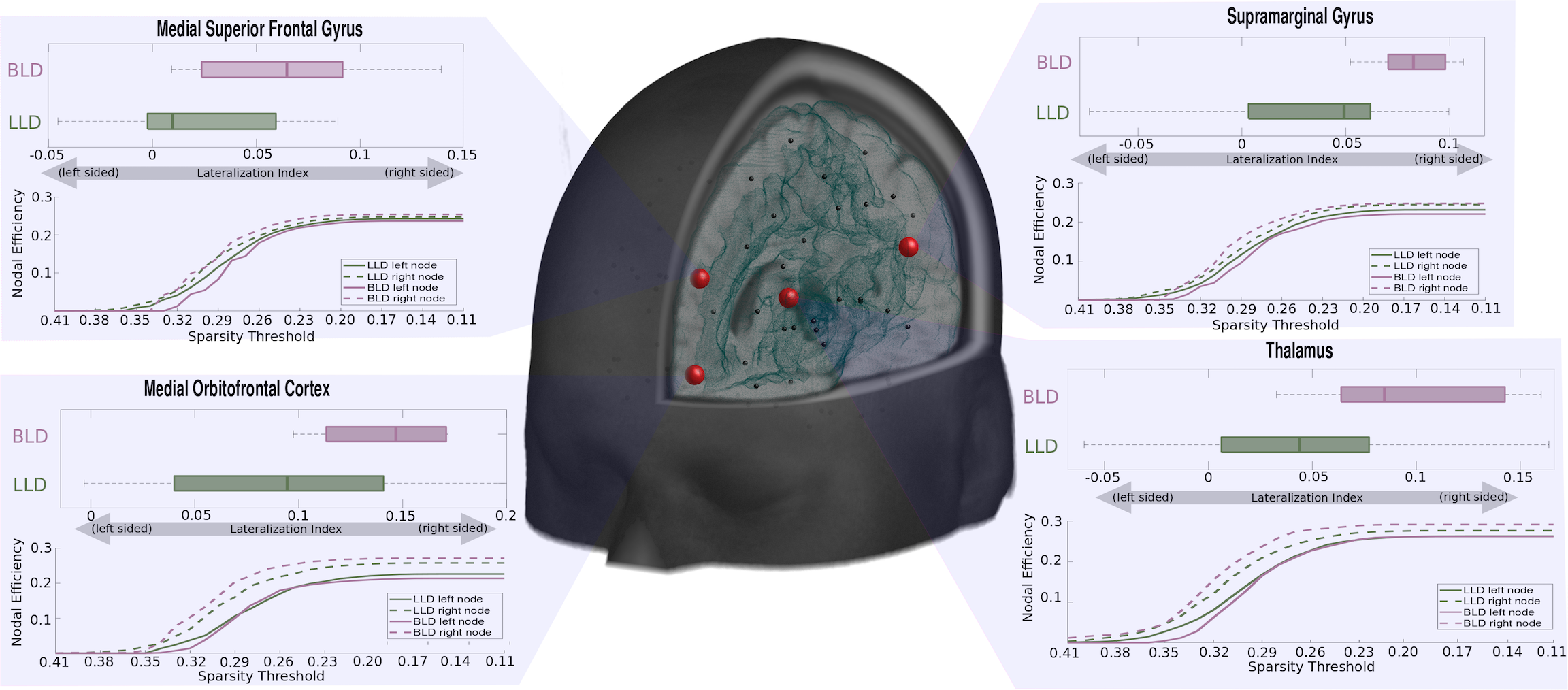
Lateralization index of regional efficiency AUC measures were significantly increased (i.e. right-sided) at 4 nodes (Fig.1) when comparing right language dominant patients to left language dominant patients, namely at the medial superior frontal gyrus (LIBLD-LLD=0.0392, p=.029), supramarginal gyrus (LIBLD-LLD=0.0467, p=.023), thalamus (LIBLD-LLD=0.0525, p=.017), and medial orbitofrontal cortex (LIBLD-LLD=0.0616, p=.026). We found that addition of these four LI values was associated with a 3% improvement to correct lateralization prediction in our hierarchical binary logistic regression model. Subsequent addition of two LI values comparing pairwise values from the medial orbitofrontal cortex to the superior temporal gyrus and the paracentral lobule similarly improved the model, with an overall model significance of .002/.007/.004, and overall correct prediction rate of 85%/88%/91%, for block 1/2/3 respectively.Discussion
Though many language-related functions lateralize in the human brain, the specific structural mapping of these networks are heterogeneous and difficult to assess with routine diffusion imaging. Nevertheless, nodal efficiency measures across multiple sparsity thresholds show promise in distinguishing bilateral language dominant and left language dominant patients, despite within-group variation in pairwise diffusion connectivity measures. Such findings may inform and constrain analysis of the data-rich structural connectome in epilepsy surgery candidates, and enhance our understanding of individualized language function. In future analyses, it may also serve as a useful starting point in better characterizing the spatial distribution and relationship between intrahemispheric and interhemispheric language networks.Conclusion
Atypical language lateralization was associated with more right-sided increases in nodal efficiencies from structural diffusion connectome data, yielding an acceptable clinical accuracy of language lateralization prediction. Future research and clinical management may benefit from an understanding of such network measures and the structural features associated with them, such that the clinical localization of language function may better match surgical structure considerations.Acknowledgements
This study was funded by grants from the National Institute of Health, R01-NS089659 to J.J. and R01-NS064033 to E.A.References
- Knecht, S. et al. Language lateralization in healthy right-handers. Brain 123, 74–81 (2000).
- Woermann, F. G. et al. Language lateralization by Wada test and fMRI in 100 patients with epilepsy. Neurology 61, 699–701 (2003).
- Möddel, G., Lineweaver, T., Schuele, S. U., Reinholz, J. & Loddenkemper, T. Atypical language lateralization in epilepsy patients. Epilepsia 50, 1505–1516 (2009).
- Rasmussen, T. & Milner, B. The role of early left-brain injury in determining lateralization of cerebral speech functions. Ann. N. Y. Acad. Sci. 299, 355–369 (1977).
- Jeong, J.-W., Asano, E., Juhász, C. & Chugani, H. T. Localization of specific language pathways using diffusion-weighted imaging tractography for presurgical planning of children with intractable epilepsy. Epilepsia 56, 49–57 (2015).
- Tailby, C., Abbott, D. F. & Jackson, G. D. The diminishing dominance of the dominant hemisphere: Language fMRI in focal epilepsy. NeuroImage Clin. 14, 141–150 (2017).
- Jeong, J.-W., Asano, E., Yeh, F.-C., Chugani, D. C. & Chugani, H. T. Independent component analysis tractography combined with a ball-stick model to isolate intravoxel crossing fibers of the corticospinal tracts in clinical diffusion MRI: New ICA+BSM Tractography Method to Isolate the Corticospinal Tracts. Magn. Reson. Med. 70, 441–453 (2013).
- Fang-Cheng Yeh, Wedeen, V. J. & Tseng, W.-Y. I. Generalized q-Sampling Imaging. IEEE Trans. Med. Imaging 29, 1626–1635 (2010).
Figures