3008
Magnetic Resonance Imaging to Determine Cerebro-Spinal Fluid Volume to Verify the Role Dehydration Plays in Traumatic Brain Injury1Physics and Mathematics, Nottingham Trent University, Nottingham, United Kingdom
Synopsis
In contact sports, it is common to see frequent head injuries or indeed traumatic brain injuries (TBI) from minor impacts which anecdotally appear increased as a result of concussion. There is little agreement in the literature regarding the change in CSF volume as a function of dehydration. Here we measure the volume using TrueFISP at 1.5T (Avanto, Siemens, DE) and thresholding images to determine the number of CSF voxels. Imaging reveals a decrease in CSF owing to dehydration. New rehydration regimens should allow for reduction in TBI incidence.
Introduction
In contact sports, it is common to see frequent head injuries or indeed traumatic brain injuries (TBI) from minor impacts. It is also common to see dehydration as players are often exerting themselves for hours with little to no rest or fluid intake. In fact in boxing and mixed martial arts (MMA) it is common practice to purposefully dehydrate oneself to reach a lower weight class. It is well accepted that due to the high specific gravity of the fluid, the Subarachnoid cerebrospinal fluid (CSF) provides a natural shock absorber to the brain, helping to prevent damage during head impacts1. CSF is created from arterial blood inside the first ventricle2 and relies on a concentration gradient between the blood and new CSF for osmosis to occur. As a direct consequence hydration level affects the rate of production of CSF and hence its total volume. The small number of publications in this area suggest that whilst the overall volume of the brain is not dependent on hydration levels3, the volume of CSF is although there is poor agreement as to whether the correlation is positive or negative4-6. If the volume of CSF decreases with dehydration the likelihood of a TBI will increase owing to reduction in the cushioning effect and thus presents opportunity to reduce TBI incidence by improving hydration. Magnetic resonance imaging (MRI) provides a valuable tool to measure the volume of the CSF during dehydration, and therefore to determine the level of protection which is afforded to the brain.Method
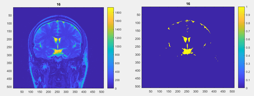
Eight preliminary participants were scanned using TrueFISP at 1.5T (Avanto, Siemens) with 5.5mm slices covering the whole skull over a 230mm FOV using a TR and TE of 4.8ms and 2.4ms respectively. Participants’ masses were measured on 3 consecutive days prior to the initial scan. Their mass was recollected immediately before the first MRI using (with Exzact EX9360 digital scales). The mass and imaging was recollected at a different time the subsequent day. Images were processed using a MATLAB (Mathworks, 2017a) script which thresholded images such that only signal from CSF and eyes remained. The number of pixels was summed and converted to approximate volume using the voxel dimensions. Interday differences as percentages were collected and plotted. The above preliminary method is then repeated with participants before and after a full rugby or football match to capture realistic levels of dehydration and CSF changes encountered during play.Results
In figure 1 a representative slice of a participant’s brain can be seen before and after processing with the MATLAB (Mathworks, 2017a) script.
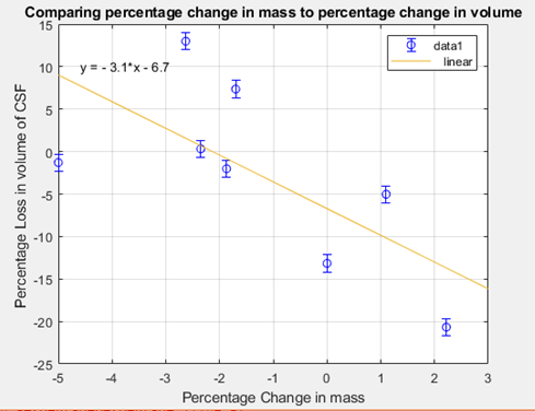
Figure 2 shows the plot of percentage mass change against percentage volume change.
Discussion
All the participants in the preliminary experiment showed a mass change higher than that of day to day fluctuations meaning it is likely they were dehydrated on one of the scan days causing the decrease/increase their body mass between scans. It is likely that hydration level is the cause of the mass changes as it is within the expected variation for this effect. A person is said to be dehydrated when they have lost 2% or more of their body mass7. Following the trend line in figure 2 it can be seen that CSF only starts to be lost after a person has lost more than 2% of their body mass, this supports the theory that dehydration is the primary cause for the loss of CSF supporting the theory that CSF volume is decreased by dehydration.Conclusion
The apparent link between reduction in CSF volume and loss of mass is likely due to dehydration. Further research is needed in order to confirm this link and validate the effect of dehydration owing to exercise. Further research will see controlled dehydration in order to confirm this if this is the reason for the link and investigating the best rehydration regimen to maintain CSF volume during high intensity exercise.Acknowledgements
The authors wish to extend their thanks to Professor Craig Sale for the initial concept for this study.References
1. Cerebrospinal Fluid (CSF). National Multiple Sclerosis Society. 2017 https://www.nationalmssociety.org/Symptoms-Diagnosis/Diagnosing-Tools/Cerebrospinal-Fluid-(CSF). Accessed November 1, 2018.
2. Welch K. Secretion of Cerebrospinal Fluid by Choroid Plexus of The Rabbit. Am J Physiol. 1963; 205:617-24.
3. Meyers, S.M. et al. J Magn Reson Imaging. 2016 Aug;44(2):296-304. doi: 10.1002/jmri.25168.
4. Dickson, J.M. et al. Int J Sports Med. 2005 Jul-Aug;26(6):481-5.
5. Curran-Sill, G. Sports Med Open. 2018 Jan 12;4(1):6. doi: 10.1186/s40798-018-0119-2.
6. Kempton, M.J. et al. Hum Brain Mapp. 2009 Jan;30(1):291-8.
7. Dehydration. nhs.uk. 2017.https://www.nhs.uk/conditions/dehydration/ Accessed November 1, 2018.
Figures