3006
Magnetic resonance elastography of repeated head impacts: Mechanical properties of the brain in collegiate hockey players1Biomedical Engineering, University of Delaware, Newark, DE, United States, 2Kinesiology and Applied Physiology, University of Delaware, Newark, DE, United States
Synopsis
In this study, we use magnetic resonance elastography (MRE) to examine the effects of a season of collegiate hockey on brain biomechanics to better understand the neurological impact of traumatic brain injury. We scanned 13 collegiate-level hockey players at four time points over the course over year using MRE to quantify the possible changes to the viscoelastic mechanical properties caused by repeated head impacts. We discovered that both stiffness and damping ratio changed over the course of the hockey season and then had some recovery after the season, indicating a complex pathology that can be quantified with MRE.
Introduction
Traumatic brain injury (TBI) presents a major problem for the healthcare industry with 1.4 million cases of TBI each year in the US [1]. Athletes in contact sports experiencing repeated, concussive and subconcussive head impacts are at heightened risk for memory dysfunction, depression, and dementia [2], though the pathological neural mechanisms that result in impairment are not well understood. Quantitative neuroimaging studies of athletes in contact sports provide a window into how the brain changes from head impacts [3]. Magnetic resonance elastography (MRE) is a novel imaging technique that can be used for noninvasively characterizing the integrity of brain tissue through mechanical properties [4,5]. In this study, we use MRE to examine the effects of a season of collegiate hockey on brain biomechanics to better understand the neurological impact of TBI.Methods
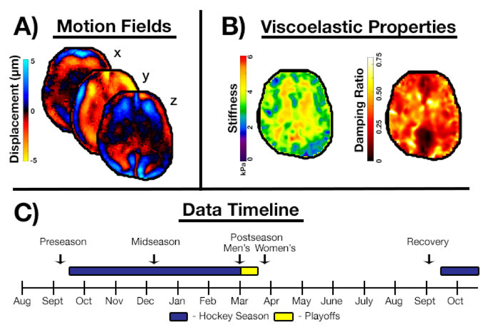
Overview of the study methods and design is shown in Figure 1.
Subjects: We examined 13 club-level college hockey players (8/5 M/F). Players were scanned four times over a year: (1) preseason, within one week prior to the first game in September 2017; (2) midseason, in December 2017 before winter break; (3) postseason, within 10 days of the last game played, in February/March 2018; and (4) recovery, before the season began in September 2018 (Figure 1C). Six players graduated and were not scanned at the recovery time point.
Imaging: All scans were performed on a Siemens 3T Prisma scanner. The scan protocol included MRE with a 3D multiband, multishot spiral sequence [6]. MRE imaging parameters included: TR/TE = 2133/70 ms; 240x240 mm2 FOV; 120x120 matrix; 64 slices; 2.0 mm isotropic resolution. Vibrations at 60 Hz were generated by the Resoundant pneumatic actuator system. Additional scans for creating anatomical regions-of-interest include diffusion tensor imaging (2.0 mm resolution, co-registered with MRE data) and 1.0 mm resolution T1-weighted MPRAGE.
Analysis: We used a nonlinear inversion algorithm (NLI) [7] to estimate the viscoelastic shear stiffness and damping ratio from measured displacement fields. The MPRAGE and DTI scans were used to create a region-of-interest incorporating all cerebral white matter (WM). We used a repeated measures ANOVA with a post-hoc Tukey test to determine whether stiffness and damping ratio changed between time points, excluding missing data and MRE scans with poor SNR [8].
Results
Stiffness was significantly different across time points (p = 0.018). Post hoc Tukey tests revealed a difference between preseason and postseason time points, with brains at postseason being 4.1% stiffer than at preseason (p = 0.015). There was also a non-significant increased stiffness at midseason and had lowered slightly at recovery, however not all the way back to preseason levels, as seen in Figure 2. Damping ratio was significantly different across time points (p = 0.005). Post hoc Tukey tests revealed differences between preseason and midseason (p = 0.042) and between midseason and recovery (p = 0.003). Brains had the lowest damping ratio at midseason (-4.1% lower than preseason), and recovered to beyond the preseason level (2.8% higher), as seen in Figure 3.Discussion and Conclusions
In this study, we observed a consistent stiffing of the brain over the course of the hockey season. This was in contrast to our initial hypothesis that softening of the brain tissue would occur due to the repeated head impacts. We also saw slight, though non-significant, recovery of the stiffness towards baseline after the season ended. These changes in stiffness may reflect increase in intracranial pressure (ICP) from the repeated head impacts during the season and a decrease in the ICP after the impacts cease, with ICP having been shown to affect MRE stiffness measures [9]. We also observed changes in the viscoelastic damping ratio during the same time span. Damping ratio decreased at the midpoint of the season, indicating a loss of microstructural integrity, and then increased at postseason and even past the baseline after recovery, indicating integrity loss is at least partially reversible [10]. Figure 4 summarizes the time course of changes in both stiffness and damping ratio. These results indicate a complex pathology caused by the repeated head impacts that can be investigated by MRE and warrants further investigation into the acute response to concussion in sports, as well as how these results correlate with other structural changes in the brain [3]. Also in future work, we will compare the MRE changes to head impact kinematics.Acknowledgements
This work was supported by the Office of Naval Research, award N00014-18-1-2018.References
[1] Langlois, J. A., Rutland-Brown, W., and Wald, M. M., 2006, The Epidemiology and Impact of Traumatic Brain Injury A Brief Overview. [
2] Baugh, C. M., Stamm, J. M., Riley, D. O., Gavett, B. E., Shenton, M. E., Lin, A., Nowinski, C. J., Cantu, R. C., McKee, A. C., and Stern, R. A., 2012, “Chronic Traumatic Encephalopathy: Neurodegeneration Following Repetitive Concussive and Subconcussive Brain Trauma,” Brain Imaging Behav., 6(2), pp. 244–254.
[3] Manning, K. Y., Schranz, A., Bartha, R., Dekaban, G. A., Barreira, C., Brown, A., Fischer, L., Asem, K., Doherty, T. J., Fraser, D. D., Holmes, J., and Menon, R. S., 2017, “Multiparametric MRI Changes Persist beyond Recovery in Concussed Adolescent Hockey Players.,” Neurology, 89(21), pp. 2157–2166.
[4] Muthupillai, R., Lomas, D. J., Rossman, P. J., Greenleaf, J. F., Manduca, A., and Ehman, R. L., 1995, “Magnetic Resonance Elastography by Direct Visualization of Propagating Acoustic Strain Waves.,” Science, 269(5232), pp. 1854–7.
[5] Hiscox, L. V, Johnson, C. L., Barnhill, E., McGarry, M. D. J., Huston, J., van Beek, E. J. R., Starr, J. M., and Roberts, N., 2016, “Magnetic Resonance Elastography (MRE) of the Human Brain: Technique, Findings and Clinical Applications,” Phys. Med. Biol., 61(24), pp. R401–R437.
[6] CL Johnson, JL Holtrop, AT Anderson, BP Sutton, “Brain MR Elastography with Multiband Excitation and Nonlinear Motion-Induced Phase Error Correction,” 24th Annual Meeting of the International Society for Magnetic Resonance in Medicine, Singapore, May 7-13, 2016, p. 1951.
[7] MDJ McGarry, EEW Van Houten, CL Johnson, JG Georgiadis, BP Sutton, JB Weaver, KD Paulsen, “Multiresolution MR Elastography Using Nonlinear Inversion,” Medical Physics, 2012; 39(10):6388-6396.
[8] McGarry, M. D. J., Van Houten, E. E. W., Perriñez, P. R., Pattison, A. J., Weaver, J. B., and Paulsen, K. D., 2011, “An Octahedral Shear Strain-Based Measure of SNR for 3D MR Elastography.,” Phys. Med. Biol., 56(13), pp. N153-64.
[9] Arani, A., Min, H.-K., Fattahi, N., Wetjen, N. M., Trzasko, J. D., Manduca, A., Jack, C. R., Lee, K. H., Ehman, R. L., and Huston, J., 2018, “Acute Pressure Changes in the Brain Are Correlated with MR Elastography Stiffness Measurements: Initial Feasibility in an in Vivo Large Animal Model,” Magn. Reson. Med., 79(2), pp. 1043–1051.
[10] Sack, I., Jöhrens, K., Würfel, J., and Braun, J., 2013, “Structure-Sensitive Elastography: On the Viscoelastic Powerlaw Behavior of in Vivo Human Tissue in Health and Disease,” Soft Matter, 9(24), p. 5672.
Figures