3005
MR Elastography (MRE)-assessed skull-brain coupling is affected by sports-related repetitive head impacts (RHI)1Radiology, Mayo Clinic, Rochester, MN, United States, 2Physiology and Biomedical Engineering, Mayo Clinic, Rochester, MN, United States, 3Naval Research Laboratory, Code 7160, Washington, DC, United States
Synopsis
Repetitive head impacts (RHI) in contact sports are known to be associated with altered brain structure and increased concussion susceptibility. Here, MR elastography-based assessment of skull-brain mechanical coupling was used as a new biomarker to assess the RHI-related injury. With novel MRE techniques to directly measure skull-brain displacement, this study aimed to determine the repeatability of MRE-measured mechanical coupling parameters and to assess their changes in RHI subjects. Results demonstrate good repeatability and show preliminary evidence that rotational transmission is significantly higher in RHI group, presumably due to the degradation of the damping capabilities of the protective pia-arachnoid complex following RHI.
Introduction
As a growing health issue, repetitive head impacts (RHI) in contact sports are known to be associated with increased concussion susceptibility, altered brain structure, and long-term neurological impairment.1-7 It is our hypothesis that repetitive sub-concussive traumatic impacts disrupt the skull-brain membrane system (known as the pia-arachnoid complex, or PAC).8,9 Despite a growing concern of the effects of RHI, identification of patients who are at risk because of prior RHI is challenging due to the lack of objective methods for assessing this condition. Here we describe our evaluation of MR Elastography (MRE)-based assessment of skull-brain coupling as a new biomarker to assess RHI-related injury to the PAC. MRE is a noninvasive imaging technique to map tissue mechanical properties.10 We have developed an MRE-based dual-saturation and dual-sensitivity motion encoding (DSDM) technique that directly measures in vivo skull-brain motion, and have studied the skull-brain coupling in volunteers with this technique.11 We proposed that if the skull-brain PAC interface was damaged during RHI, mechanical tethering between the skull and brain would be affected, and the MRE-assessed skull-brain coupling behavior would be changed following RHI. This preliminary study thus aimed to determine the repeatability of DSDM-MRE-assessed skull-brain coupling parameters and to assess their changes in RHI subjects.Methods
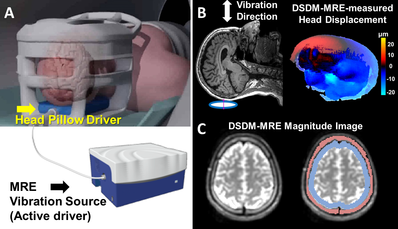
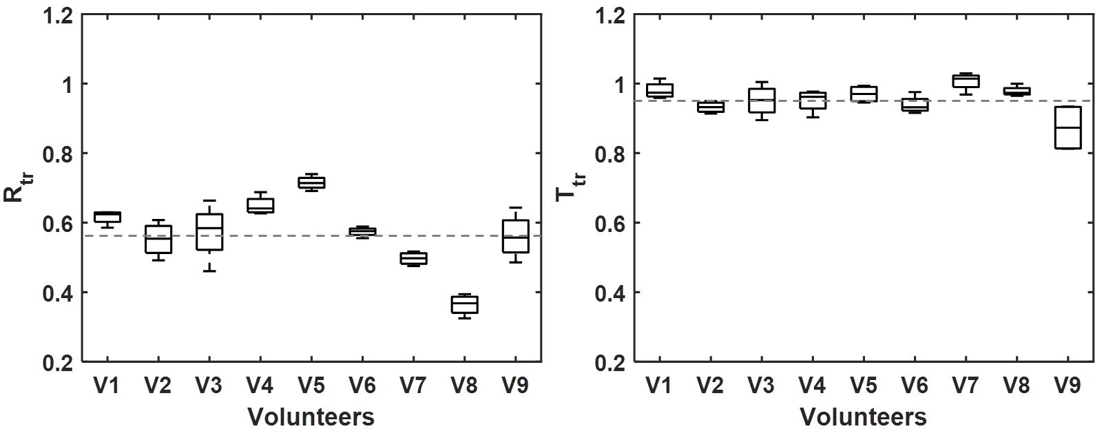
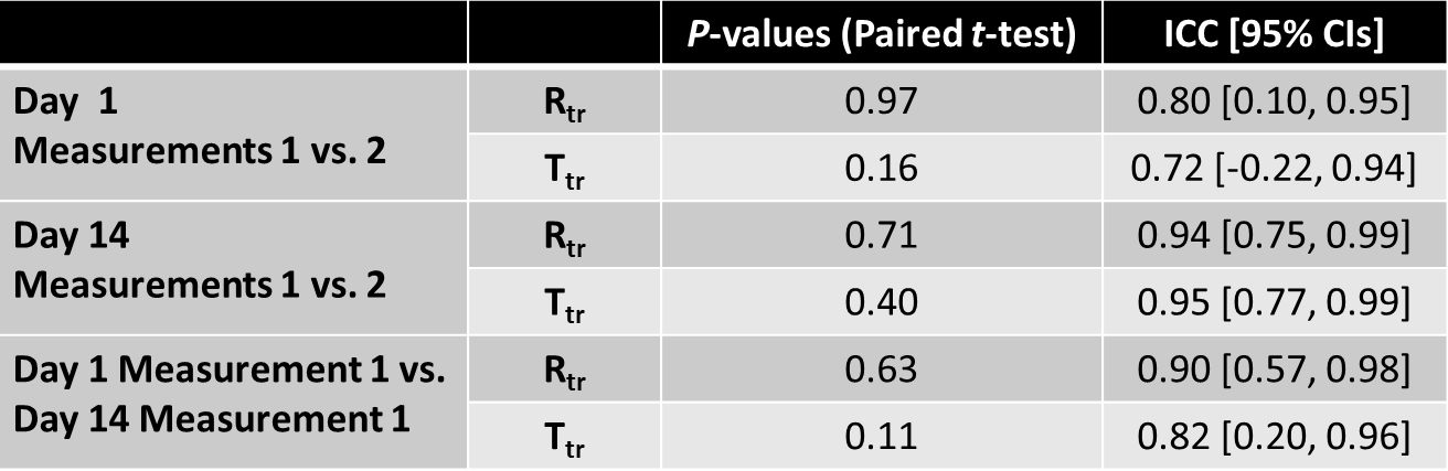
After institutional IRB approval and written informed consent, 9 healthy volunteers (age 43±18yrs) with no history of contact-sports participation (RHI-) and 5 ice hockey players (age 18±1yrs) with ~6-16 years participation of hockey (RHI+) but with no history of severe traumatic brain injury were assessed with DSDM-MRE on a 3T head-only MR scanner.12 Briefly, low amplitude 60-Hz mechanical vibration was introduced from the skull to the brain via a soft pillow-like driver placed under the head (Figure-1). The resulting 3D full-volume head displacement field was imaged by DSDM-MRE as previously described.11 The repeatability study was only performed on volunteers, with DSDM-MRE performed twice on day-1 (separated by a 5-minute break leaving the scanner) and repeated after 14 days. MRE displacements from the skull/brain ROIs were fitted to a model of rigid-body motion to obtain rigid-body rotation and translation. Rotational and translational transmission ratio Rtr and Ttr (defined as the brain-to-skull amplitude ratio of the rotation and translation, respectively) was calculated to characterize the skull-brain coupling. The within-day and between-day repeatability were analyzed with intraclass correlation coefficients (ICCs). A paired t-test was used to identify differences between the tests and retests. The Mann-Whitney U test was used to measure the differences between mean Rtr and Ttr values of the RHI- and RHI+ groups. P-value<0.05 was defined as significant.Results
Figure-2 displays measured Rtr and Ttr values from the 4 DSDM-MRE scans for all 9 volunteers. The paired t-test results did not show any significant difference between the pairs of measurements obtained on the same day or separate days. The Rtr and Ttr showed good-to-excellent repeatability (ICCs:0.72-0.95) for within- and between-day measurements (Table-1). Figure-3 compares the measured Rtr and Ttr between RHI- and RHI+ groups. We found significantly elevated Rtr in ice hockey players (RHI+) compared with 9 healthy controls, but no significance was found for Ttr.Discussion
Our results demonstrate good overall repeatability of DSDM-MRE-assessed skull-brain coupling parameters, which suggests high reliability for our approach. The skull-brain motion analysis in volunteers also demonstrated that the skull-brain interface significantly attenuates the rotational motion (Rtr=0.57±0.10) that is potentially more harmful in regard to brain injury than translational motion (Ttr=0.95±0.05). This agrees with other studies suggesting that the skull-brain membrane system plays an important role in damping and constraining the brain motion relative to the skull under an impact.13-18 Based on our hypothesis, when the head was vibrated, mechanical interactions between the skull-brain interface, especially meninges, cerebrospinal fluid, arachnoid trabeculae, and subarachnoid vasculature provide a hydro-mechanical shock absorber system that attenuates the motion transmission from the skull to the brain. However, if the tethering structure is injured, such attenuation effects would be degraded, as demonstrated by increased Rtr in the RHI+ group. However, noting the age difference between the RHI- and RHI+ groups in this preliminary study, our future work will focus on recruiting more subjects to perform the age-matched comparison to further validate our findings.Conclusion
The results show that MRE-based techniques can be used to characterize the functional status of the PAC in protecting the brain from mechanical transients applied to the skull. Our results also provide preliminary evidence that individuals who have previously experienced RHI have changes in skull-brain coupling characteristics that may represent the degradation of the protective function of PAC, thereby placing them at greater risk for future injury.Acknowledgements
This study is funded by NIH Grants RO1 EB001981, RO1 EB010065, and Office of Naval Research Contract N00014-18-C-2016.References
1. Mainwaring, L, Ferdinand Pennock, KM, Mylabathula, S, et al. Subconcussive head impacts in sport: A systematic review of the evidence. Int J Psychophysiol. 2018;132(Pt A):39-54.
2. Bailes, JE, Petraglia, AL, Omalu, BI, et al. Role of subconcussion in repetitive mild traumatic brain injury. J Neurosurg. 2013;119:1235-1245.
3. Beckwith, JG, Greenwald RM, Chu JJ, et al. Head impact exposure sustained by football players on days of diagnosed concussion. Med Sci Sports Exerc. 2013;45(4):737-46.
4. Guskiewicz, KM, McCrea M, Marshall SW, et al. Cumulative effects associated with recurrent concussion in collegiate football players: the NCAA Concussion Study. JAMA. 2003;290(19):2549-2555.
5. Talavage, TM, Nauman EA, Breedlove EL, et al. Functionally-detected cognitive impairment in high school football players without clinically-diagnosed concussion. J Neurotrauma. 2014;31(4):327-338.
6. Lipton, ML, Kim N, Zimmerman ME, et al. Soccer Heading Is Associated with White Matter Microstructural and Cognitive Abnormalities. Radiology. 2013:268(3): 850-857.
7. Montenigro, PH, Alosco ML, Martin BM, et al. Cumulative Head Impact Exposure Predicts Later-Life Depression, Apathy, Executive Dysfunction, and Cognitive Impairment in Former High School and College Football Players. J Neurotrauma. 2017;34(2):328-340.
8. Scott, G, Coats, B. Microstructural characterization of the pia-arachnoid complex using optical coherence tomography. IEEE Trans Med Imaging. 2015;34(7):1452-1459.
9. Saboori, P, Sadegh, A. Histology and morphology of the brain subarachnoid trabeculae. Anat Res Int. 2015;2015:279814.
10. Yin, Z, Romano, AJ, Manduca, A, et al. Stiffness and Beyond: What MR Elastography Can Tell Us About Brain Structure and Function Under Physiologic and Pathologic Conditions. Top Magn Reson Imaging. 2018;27(5):305-318.
11. Yin, Z, Sui Y, Trzasko JD, et al. In vivo characterization of 3D skull and brain motion during dynamic head vibration using magnetic resonance elastography. Magn Reson Med. 2018;doi: 10.1002/mrm.27347.
12. Weavers, PT, Shu Y, Tao S, et al. Technical Note: Compact three-tesla magnetic resonance imager with high-performance gradients passes ACR image quality and acoustic noise tests. Med Phys. 2016;43(3):1259-64.
13. Hardy, WN, Foster CD, Mason MJ, et al. Investigation of Head Injury Mechanisms Using Neutral Density Technology and High-Speed Biplanar X-ray. Stapp Car Crash J. 2001;45:337-68.
14. Zou, H, Schmiedeler, JP, Hardy, WN. Separating brain motion into rigid body displacement and deformation under low-severity impacts. J Biomech. 2007;40(6):1183-91.
15. Ji, S, Roberts, DW, Hartov, A, et al. Brain-skull contact boundary conditions in an inverse computational deformation model. Send toMed Image Anal. 2009;13(4):659-72.
16. Bayly, PV, Cohen TS, Leister EP, et al. Deformation of the human brain induced by mild acceleration. J Neurotrauma. 2005;22(8):845-56.
17. Clayton, EH, Genin, GM, Bayly, PV. Transmission, attenuation and reflection of shear waves in the human brain. J R Soc Interface. 2012;9(76):2899–2910.
18. Badachhape, AA, Okamoto RJ, Durham RS, et al. The relationship of three-dimensional human skull motion to brain tissue deformation in magnetic resonance elastography studies. J Biomech Eng. 2017;139(5). doi: 10.1115/1.4036146.
Figures