3003
Decreased Brain Temperature in Former NFL Athletes1Radiology, Brigham and Women's Hospital, Boston, MA, United States, 2Center For Clinical Spectroscopy, Brigham and Women's Hospital, Boston, MA, United States, 3Boston University, Boston, MA, United States, 4Psychiatry Neuroimaging Laboratory, Brigham and Women's Hospital, Boston, MA, United States, 5Psychiatry, Massachusetts General Hospital, Boston, MA, United States, 6Child and Adolescent Psychiatry, Ludwig-Maximilians-Universität, Munich, Germany, 7VA Boston Healthcare System, Brockton, MA, United States
Synopsis
Currently, Chronic Traumatic Encephalopathy (CTE) is only diagnosed post-mortem, therefore advanced imaging has an opportunity to identify biomarkers for this disease. This study’s goal is to use Magnetic Resonance Spectroscopy (MRS) and MR Thermography (MRT) to measure cerebral temperature differences between retired former NFL players (n = 50) suspected of CTE and controls (n = 13). The NFL players were found to have lower brain temperature than the controls (p = 0.0340). These finding suggest there is a metabolic difference between those suspected of CTE and healthy controls.
Introduction
Repetitive mild traumatic brain injury (rmTBI) in contact sports, such as tackle football, can lead to long-term cognitive and neuropsychiatric deficits. One disease that is heavily linked with rmTBI is chronic traumatic encephalopathy (CTE) - a neurodegenerative disease that is associated with impaired cognition, chronic headache, and cerebellar dysfunction [1]. CTE is currently only diagnosed post-mortem, and there is a need to detect this neurodegenerative disease in vivo– potentially through radiology imaging. While some advanced imaging modalities focus on structural changes, Magnetic Resonance Spectroscopy (MRS) allows us to monitor the neurochemical changes following injury and in neurodegeneration, which may be helpful in the diagnosis of CTE in the future. Additionally, MRS can be used for MR Thermography (MRT), a non-invasive, in vivotemperature measurement using the temperature dependent chemical shift values.In this study, we compared MRS and MRT results in a sample of symptomatic former NFL players with same-age, asymptomatic controls (without contact sport experience or TBI history) to examine metabolite concentration and brain temperature differences.Methods
MRS single voxel PRESS scans were performed on a 3T Siemens scanner (2x2x2 cm3voxel size, TE: 30ms, TR: 2000ms, 128 repetitions) on a subset subjects in the NIH-funded DETECT study: 50 former NFL players (ages 40-69) and 13 controls. Scans were performed in the posterior cingulate gyrus (PCG) and the parietal white matter (PWM). MRS data was processed using OpenMRSLab and LC Model. MRT data was processed using OpenMRSLab in combination with the PeakUtils python library. This software determines the frequency of water (which is temperature dependent) and the frequency of N-acetyl aspartate (which is temperature independent) and calculates the difference between those frequency values. Calibration scans were conducted and showed that a larger PPM difference correlates with a lower temperature. In addition, T1 weighted images were acquired and whole brain volume was measured using an FSL mask.Results
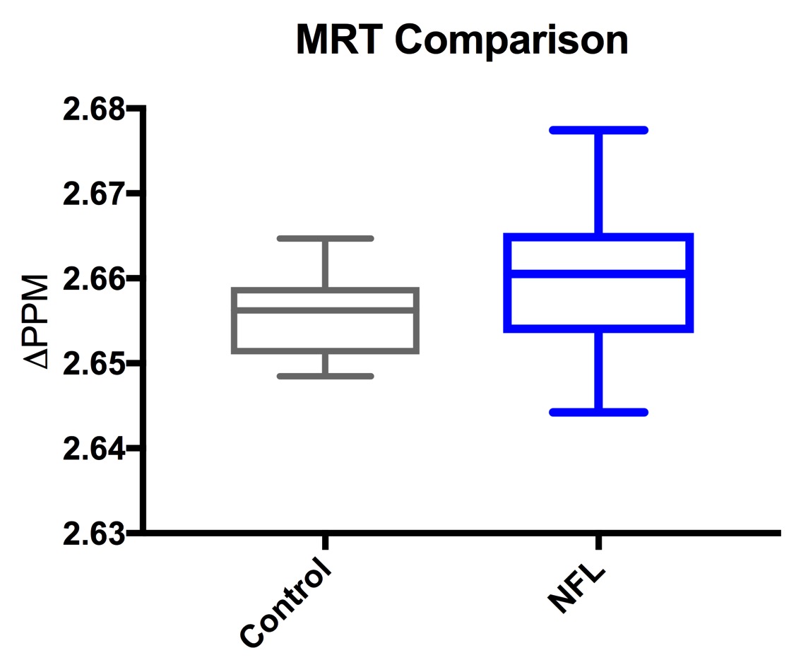
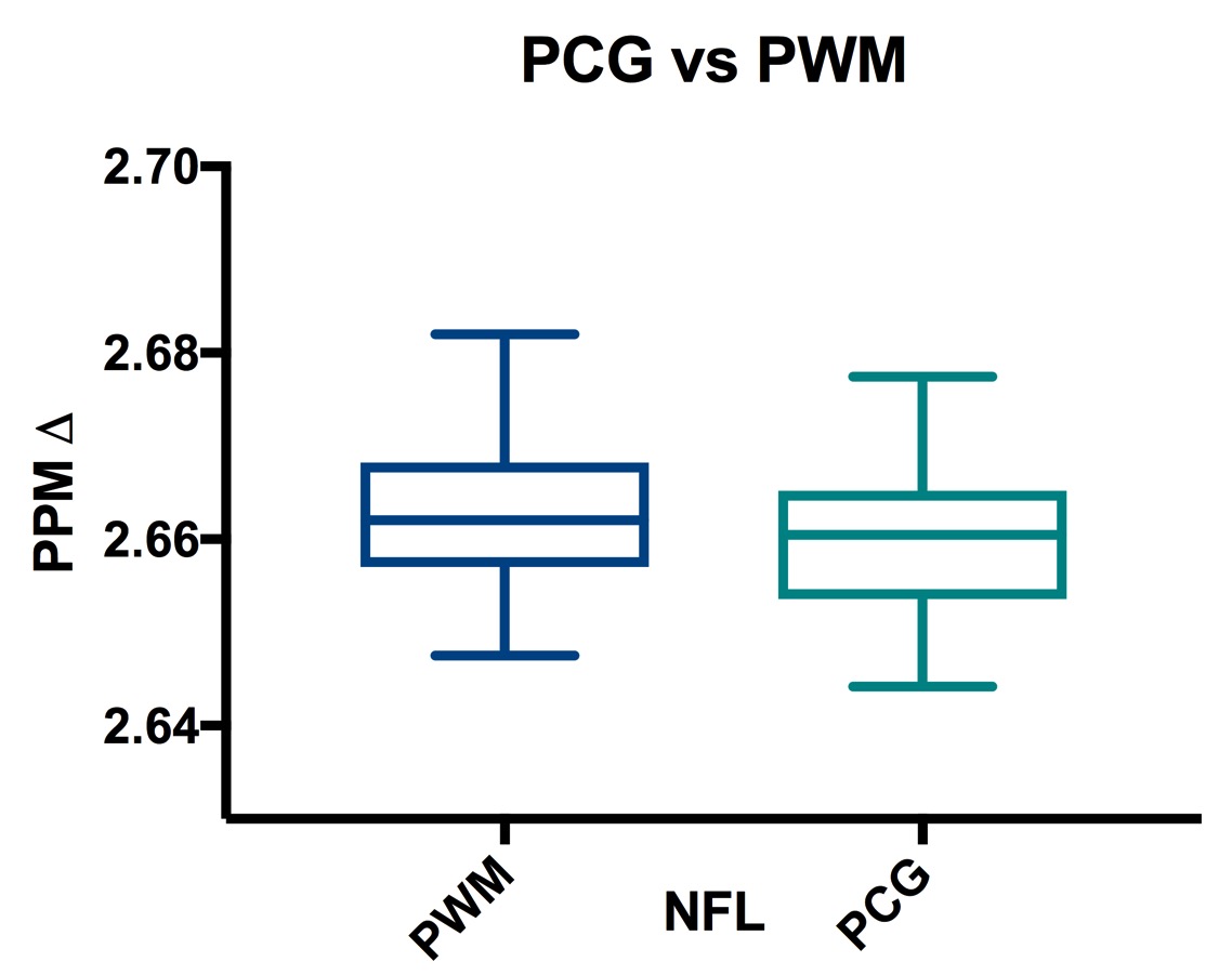
For the PCG, the NFL and control groups differed for the ΔH2O-NAA values (p=0.0340) (Figure 1). The NFL group was found to have a higher PPM difference, which correlates with a lower temperature, as found in earlier calibration scans. For the PWM, there was no significant difference in MRT values. When comparing the PCG and PWM MRT values, the two regions were found to be statistically different via a paired T Test for the NFL group (p=0.0411) (Figure 2). Also, the PCG and PWM MRT values were significantly positively correlated (p=0.0023, r=0.4055). In the PCG, ΔH2O-NAA was significantly correlated with glutathione (GSH) over total creatine (GSH/tCr) (p=0.0006, r= 0.47) (Figure 3). Additionally, there was no difference in the brain volume of the two groups, and nor was there a correlation between brain volume and ΔH2O-NAA.Discussion
An increased brain temperature could be linked to dysfunctional metabolism or inflammation while a decrease in brain temperature may be associated with a decrease in cerebral brain flow (CBF) [2]. Reduced CBF has been found in former NFL players [3], and Ojo et al (2016) also saw reduced CBF in mice following rmTBI [4]. This decrease in brain temperature may be related to a decrease in brain metabolism, which is tightly linked to brain temperature [2]. It is particularly interesting that GSH/tCr and ΔH2O-NAA are positively correlated. GSH decreases with inflammation and brain temperature increases with inflammation. However, since ΔH2O-NAA is negatively correlated with brain temperature, the correlation between the temperature and metabolite measurement both support the presence of inflammation in these NFL athletes. Furthermore, the difference between the PCG and PWM is also interesting and suggests that there may be a metabolic activity difference between the two areas. The lack of differences in brain volume and correlation with brain temperature show that size of the subjects was not a confounding factor.The exact brain pathology and relationship with brain temperature should be investigated further to see how brain temperature can be better used as a biomarker in TBI research.Acknowledgements
We would like to acknowledge funding from the following grants: R01AG038758-01 and 1U01NS093334-01References
1. Stern R. et al. “Clinical presentation of chronic traumatic encephalopathy.” Neurology. 2013; 81:1122-1129.
2. Mrozek S, Vardon F, Geeraerts, T. “Brain Temperature: Physiology and Pathophysiology after Brain Injury.” Anestheiology Research and Practice. 2012.
3. Amen DG, et al. Perfusion Neuroimaging Abnormalities Alone Distinguish National Football League Players from a Healthy Population. J Alzheimers Dis 2016; 53:237-41.
4. Ojo JO, et al. Chronic Repetitive Mild Traumatic Brain Injury Results in Reduced Cerebral Blood Flow, Axonal Injury, Gliosis, and Increased T-Tau and Tau Oligomers. Journal of neuropathology and experimental neurology 2016;75:635-55.
Figures