2999
Significant Reductions in Brain Cortical Volumes and Regional Cerebral Blood Flows after Playing College Football 2 to 3 Years1Department of Radiology, Michigan State University, East Lansing, MI, United States, 2Department of Intercollegiate Athletics, Michigan State University, East Lansing, MI, United States, 3Department of Neurology and Ophthalmology, Michigan State University, East Lansing, MI, United States
Synopsis
There has been growing concern over sports-related brain injuries and their long-term effects. However, the cumulative effect of sub-concussive hits on the brain is still poorly understood. Twenty-one male Division I collegiate football athletes completed T1 volumetric and arterial spin labeling MRI scans at freshman year with follow-up 2-3 years later. Significant reductions in both brain global and regional cortical volumes were observed. Interestingly, cerebral blood flow was significantly reduced in regions associated with the default-mode network. These changes point to potential long-term effects of sub-concussive hits on the brain.
INTRODUCTION
There has been growing concern over sports-related brain injuries and their long-term effects (1). The cumulative effect of sub-concussive hits on the brain is still poorly understood. Collegiate football athletes, who frequently encounter sub-concussive hits, are an ideal population to address this question. In a recent study, alterations in functional connectivity and cerebral blood flow (CBF) in collegiate football players were found over a single football season (2). To investigate further, we conducted a study with 2-3 year follow-up to examine long-term changes.METHODS
Acquisition: Twenty-one male Division I collegiate football athletes participated in this study. They completed MRI scans at freshman year as baseline and were followed up 2-3 years later. MRI images were collected on a GE 3T Signa® HDx MR scanner with an 8-channel head coil. High-resolution 3D IR FSPGR T1-weighted images were collected with: 180 1-mm sagittal slices, CSF suppressed, TE = 3.8ms, TR of acquisition = 8.6ms, TI = 831ms, TR of inversion = 2332ms, flip angle = 8°, FOV = 25.6cm×25.6 cm, matrix size = 256×256, and receiver bandwidth = ± 20.8kHz. Then 3D pseudo continuous ASL images were collected on 12 of these athletes to measure regional CBF with: fast spiral acquisition, 32 4-mm axial slices, TE = 9.8ms, TR = 4.56s, effective resolution = 3.22mm×3.22mm, reconstructed matrix size = 128×128, NEX = 3, FOV = 22cm×22cm, receiver bandwidth = ± 62.5 kHz, labeling duration = 1.45s, saturation time = 2s, post labeling delay = 1.525s.
Volumetric analyses: Brain segmentation was conducted with FreeeSurfer (3). Two-tail paired t-tests were conducted on all brain volumes extracted over two time points, including global and regional cortical, sub-cortical and global white-matter volumes. Bonferroni correction was applied to correct the 104 comparisons. P value significance was set at ≤ 0.05 after, or ≤ 4.81 × 10⁻⁴ before correction.
CBF analyses: The CBF mean value was calculated for each of the 86 cortical and subcortical regions based on FreeSurfer segmentation (3). Two-tail paired t-tests were applied to the mean CBF values at these regions to assess the difference between the baseline and follow-up scans. P value significance was set at ≤ 0.05 after, or ≤ 5.88 × 10⁻⁴ before correction.
RESULTS
Volumetric analyses: There was a significant 1.53% reduction in global brain volume, excluding ventricles. A 2.71% reduction of total gray-matter volume contributed to this change. No changes were observed in total cerebral white-matter volume. Additionally, a 3.54% and 3.45% reduction of the right and left cortical volumes respectively contributed to the loss in total gray-matter volume. The total sub-cortical volume appeared unchanged. Significant volume reductions were found in 20 cortical regions dispersed across all four lobes and one sub-cortical region.
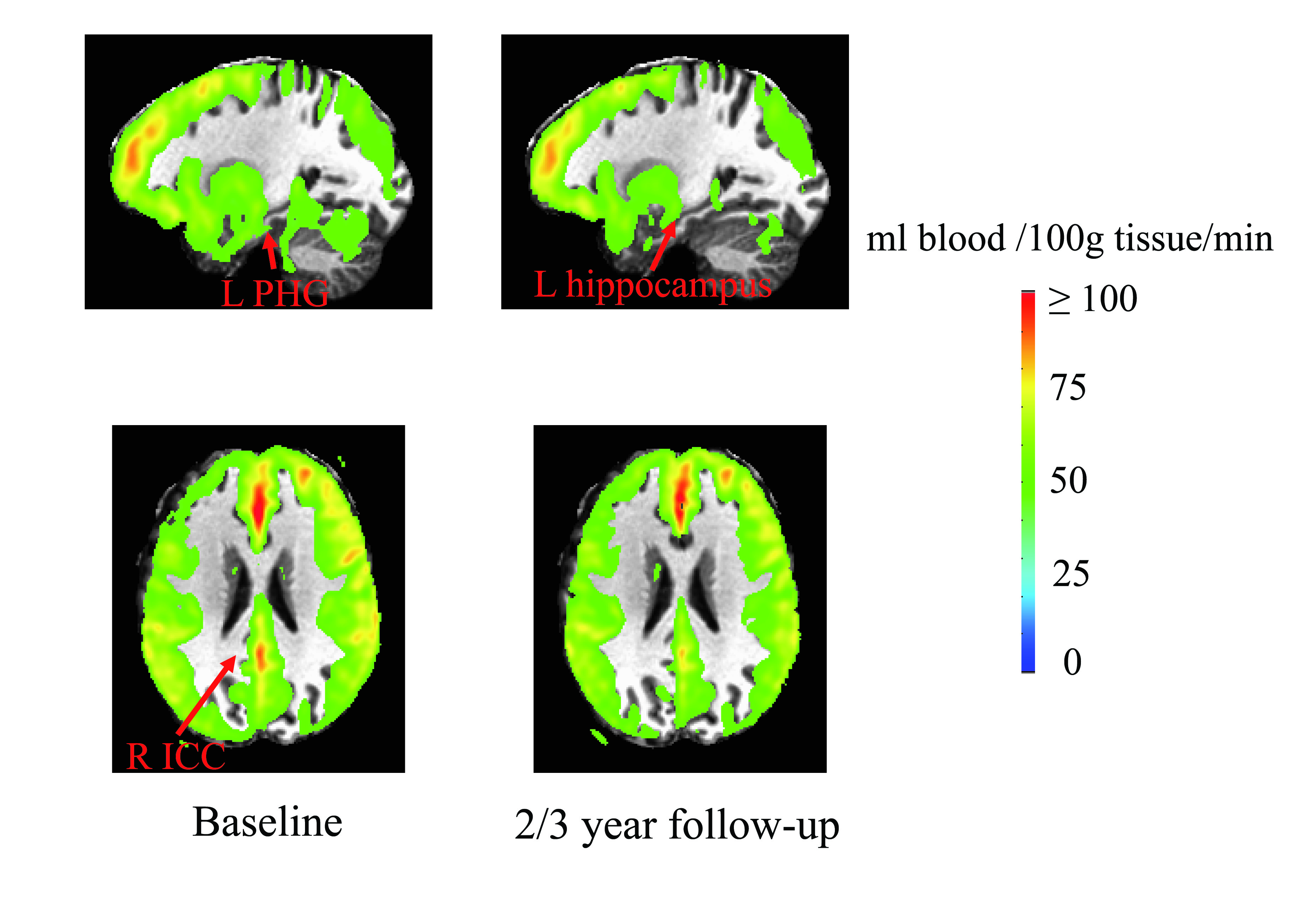
CBF analyses: CBF values were significantly reduced from baseline to follow-up at the right isthmus of the cingulate cortex (ICC) (59.8±5.9 to 51.7±7.6 ml blood/100g tissue/min, P = 0.0277), left parahippocampal gyrus (PHG) (41.5±5.9 to 31.8±6.8 ml/100g tissue/min, P = 0.0013), left medial orbitofrontal cortex (MeOFC) (56.5±10.8 to 38.8±7.0 ml/100g tissue/min, P = 0.0074), left fusiform gyrus (44.4±6.2 to 36.1±5.5 ml/100g tissue/min, P = 0.014) and left hippocampus (45.3±4.6 to 39.0±6.2 ml/100g tissue/min, P = 0.0175) (Fig. 1).
DISCUSSION AND CONCLUSION
Over only a single football season, significant changes were found in functional connectivity and CBF (2), while the brain structure remained stable. The brain structure remained stable even 30 days after a concussion (4). It is reasonable to speculate that the many repeated sub-concussive hits occurring between baseline and 2-3 year follow-up resulted in subtle changes in brain structure and micro vasculature, which eventually led to neuronal damage resulting in the observed reduction of cortical volume. The brain cortical volume reduction appeared to disperse over all four lobes of the brain. This lack of anatomical specificity might reflect the multi-directional head impacts occurring in a football. Prior studies demonstrated that the default-mode network (DMN) connectivity was altered due to concussion (4) and sub-concussive hits (2). ICC is within the hub region of DMN. MeOFC, PHG and hippocampus are also nodes in the DMN. Brain damages at various locations might have led to the reduction of functional interaction to and within the DMN, and thus led to reduction of metabolic activity, reflected by CBF, at the DMN nodes. These changes necessitate future studies on how sub-concussive hits effect brain structure, networks, metabolic activity and vasculature longitudinally.Acknowledgements
No acknowledgement found.References
1. McKee AC, Cantu RC, Nowinski CJ, Hedley-Whyte ET, Gavett BE, Budson AE, Santini VE, Lee HS, Kubilus CA, Stern RA. Chronic traumatic encephalopathy in athletes: progressive tauopathy after repetitive head injury. J Neuropathol Exp Neurol 2009;68(7):709-735.
2. Slobounov SM, Walter A, Breiter HC, Zhu DC, Bai X, Bream T, Seidenberg P, Mao X, Johnson B, Talavage TM. The effect of repetitive subconcussive collisions on brain integrity in collegiate football players over a single football season: A multi-modal neuroimaging study. Neuroimage Clin 2017;14:708-718.
3. Fischl B, Salat DH, Busa E, Albert M, Dieterich M, Haselgrove C, van der Kouwe A, Killiany R, Kennedy D, Klaveness S, Montillo A, Makris N, Rosen B, Dale AM. Whole brain segmentation: automated labeling of neuroanatomical structures in the human brain. Neuron 2002;33(3):341-355.
4. Zhu DC, Covassin T, Nogle S, Doyle S, Russell D, Pearson RL, Monroe J, Liszewski CM, DeMarco JK, Kaufman DI. A potential biomarker in sports-related concussion: brain functional connectivity alteration of the default-mode network measured with longitudinal resting-state fMRI over thirty days. J Neurotrauma 2015;32(5):327-341.
Figures