2998
High impact sports and microstructural changes in cortical brain tissue: a 4-year longitudinal study of collegiate athletes1Stanford, Stanford, CA, United States, 2University Medical Center Hamburg-Eppendorf, Hamburg, Germany
Synopsis
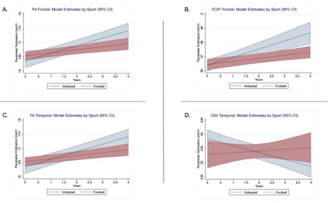
Exposure to repeated high-velocity impacts may contribute to increased risk of cognitive impairment. However, there has been limited long-term longitudinal investigations into brain tissue change in high-impact sports. In this large 4-year longitudinal DTI study, high (football) and low-contact (volleyball) athletes show a temporal double dissociation in cortical microstructure: in both the frontal and temporal lobes, cortical FA increases over time in volleyball compared to football. While an increase in ICVF underlies this FA increase in frontal cortex, a decrease in ODI underlies this FA increase in temporal cortex. Exposure to high-impact sports may alter cortical microstructural development.
Introduction
Repeated concussive and sub-concussive high-velocity impacts may contribute to brain injury and cognitive impairment in athletes of high-contact sports such as football1–3. Noninvasive brain imaging can potentially characterize the subtle changes that occur with symptomatic mild traumatic brain injury, which can have long-term consequences. Recent cross-sectional MR imaging studies have demonstrated that repetitive concussive and sub-concussive injury in collegiate football athletes is associated with brain changes compared to healthy controls4. Such findings are especially relevant because long term consequence of repeated concussive and sub-concussive impacts in contact sports, such as football, may impact brain health into adulthood, increase risk of neurodegenerative and psychiatric disease, and in severe cases result in chronic traumatic encephalopathy (CTE)5–7, a serious neurodegenerative disease found in athletes engaging in high impact sports. The current work investigates how exposure to high impact sports relates to brain change in colligate athletes. Specifically, we investigate longitudinal changes in diffusion tensor imaging (DTI) derived measures of brain tissue microstructure resulting from collegiate football.Methods
We prospectively enrolled 63 high-contact (football) collegiate athletes and 34 low-contact (volleyball) collegiate athletes over the course of 4 years. Subjects were scanned yearly and after concussion. In total, 181 football and 81 volleyball scans were analyzed (all males). Using a 3T scanner (GE MR 750) and an 8-channel receive head coil, we acquired 1.875x1.875x2.0mm whole-brain DTI with 60/30 directions at b=2500/800 s/mm2 and 9 b=0s/mm2 images. A field-map was used to correct distortions, and eddy/motion correction was performed with FSL 5.0.12. Regions of interest (ROI) were defined based on the cortical parcellation from a Freesurfer longitudinal pipeline8,9. These ROIs were combined based on anatomical hierarchy resulting in 14 bilateral regions: Caudate, Putamen, Pallidum, Hippocampus, Thalamus, Amygdala, Corpus callosum, Temporal lobe, Cingulate, Insula, Frontal lobe, Occipital lobe, Parietal lobe, subcortical white matter, and deep white matter. The following diffusion tensor imaging (DTI) metrics were used: fractional anisotropy (FA), mean diffusivity (MD) as well as Neurite Orientation Dispersion and Density Imaging (NODDI)10 derived metrics of orientation dispersion (ODI), intra-cellular volume fraction (ICVF), and the free water volume fraction (FISO). A linear mixed effects model examined changes the effect of sport (football versus volleyball), age at time of baseline scanning, years after baseline scan (time), and the interaction between time and sport. Bonferroni correction was based on 14 ROIs.Results
Different trajectories in tissue microstructure were found in football compared to volleyball athletes in both the frontal and temporal lobes with a significant time by sport interaction. In the temporal lobe, FA increases more with time in volleyball compared to football (interaction raw p = .0024, corrected p = .033, t = -3.06). This is accompanied by a decrease with in age in ODI in volleyball but not football athletes (interaction: raw p = .0002, corrected p = .003, t = -3.68). In the frontal lobe, the same trajectory is found where FA increases more quickly in volleyball athletes (interaction raw p = .003, corrected p = .045, t = -2.97). This is accompanied by a similar change in ICVF where volleyball athletes show a greater increase in ICVF with time compared to football athletes (interaction raw p = .001, corrected p = .024, t = -3.16). No significant change was found in MD or FISO. No other regions show significant change after Bonferroni adjustment.Discussion
Over the course four years, football athletes show differential trajectories in brain tissue microstructure compared to volleyball athletes. Specifically, the frontal cortical FA increase is associated with increased ICVF, that could speculatively be caused by a strengthening of the normal radial fibers in the cortex. In contradistinction, the temporal cortical FA increase is associated with decreased ODI, suggesting a loss of tangential cortical fibers.Conclusion
Exposure to repeated head impact and sub-concussive injury may play a role in altering the developmental trajectory of cortical tissue microstructure. Future work will be needed in order to fully understand if impact severity directly relates to changes in these tissue properties.Acknowledgements
No acknowledgement found.References
1. Weinberger, B. C. & Briskin, S. M. Sports-Related Concussion. Clin. Pediatr. Emerg. Med. 14, 246–254 (2013).
2. Bailes, J. E., Petraglia, A. L., Omalu, B. I., Nauman, E. & Talavage, T. Role of subconcussion in repetitive mild traumatic brain injury. J. Neurosurg. (2013). doi:10.3171/2013.7.JNS121822
3. DeKosky, S. T., Ikonomovic, M. D. & Gandy, S. Traumatic Brain Injury — Football, Warfare, and Long-Term Effects. N. Engl. J. Med. (2010). doi:10.1056/NEJMp1007051
4. Davenport, E. M. et al. Abnormal White Matter Integrity Related to Head Impact Exposure in a Season of High School Varsity Football. J. Neurotrauma (2014). doi:10.1089/neu.2013.3233
5. McKee, A. C. et al. Chronic traumatic encephalopathy in athletes: Progressive tauopathy after repetitive head injury. Journal of Neuropathology and Experimental Neurology (2009). doi:10.1097/NEN.0b013e3181a9d503
6. Gavett, B. E., Stern, R. A. & McKee, A. C. Chronic Traumatic Encephalopathy: A Potential Late Effect of Sport-Related Concussive and Subconcussive Head Trauma. Clinics in Sports Medicine (2011). doi:10.1016/j.csm.2010.09.007
7. Omalu, B. et al. Emerging histomorphologic phenotypes of chronic traumatic encephalopathy in american athletes. Neurosurgery (2011). doi:10.1227/NEU.0b013e318212bc7b
8. Fischl, B. FreeSurfer. NeuroImage (2012). doi:10.1016/j.neuroimage.2012.01.021
9. Reuter, M., Rosas, H. D. & Fischl, B. Longitudinal FreeSurfer for Reliable Imaging Biomarkers. Proc. MICCAI Nov. Neuroimaging Biomarkers Alzheimer’s Dis. Relat. Disord. Work. Chall. (2012).
10. Zhang, H., Schneider, T., Wheeler-Kingshott, C. A. & Alexander, D. C. NODDI: Practical in vivo neurite orientation dispersion and density imaging of the human brain. Neuroimage (2012). doi:10.1016/j.neuroimage.2012.03.072
Figures