2996
Structural and functional neuroimaging changes in female rugby players with and without a history of concussion1Radiology, University of Calgary, Calgary, AB, Canada, 2Kinesiology, Western University, London, ON, Canada, 3Primary Care Sport Medicine, Fowler Kennedy Sports Medicine, London, ON, Canada, 4Molecular Medicine, Robarts Research Institute, London, ON, Canada, 5Health and Rehabilitation Sciences, Western University, London, ON, Canada, 6Anatomy and Cell Biology, Western University, London, ON, Canada, 7Medical Biophysics, Western University, London, ON, Canada, 8Physical Medicine and Rehabilitation, Western University, London, ON, Canada, 9Paediatrics Critical Care Medicine, Western University, London, ON, Canada, 10Occupational Therapy, Western University, London, ON, Canada, 11Microbiology and Immunology, Western University, London, ON, Canada, 12Centre for Functional and Metabolic Mapping, Robarts Research Institute, London, ON, Canada
Synopsis
In this study we acquired diffusion and resting state fMRI data from female varsity rugby players, rowers and swimmers during the in- and off-season and found (a) significant alterations in the corpus callosum that correlated with altered default mode network connectivity with the posterior cingulate cortex as well as (b) fluctuations in white matter diffusion measures within the brainstem in contact athletes compared to non-contact athletes. Together this suggests that repetitive subclinical impacts incur both acute and long-term changes to brain microstructure and function despite lack of symptoms or even a history of concussion.
Introduction
Athletes engaged in contact sports experience repetitive subclinical impacts. Recent studies suggest that these seemingly innocuous impacts could have a significant cumulative effect on brain health (1-3). Non-invasive neuroimaging techniques sensitive to brain microstructure and function have been used to demonstrate alterations in contact athletes that correlate with the number of subclinical impacts received during a season of play (4,5). However it is essential to understand the evolving nature of these changes throughout single or multiple seasons of contact play relative to non-contact athletes and the influence of a concussion history on these brain changes.Methods
We followed a women’s varsity rugby team (contact) over multiple seasons (n = 70) and compared them to a single season of age-matched female varsity rowers and swimmers (n = 31, non-contact). These athletes did not experience a concussion within 6-months of data collection and a sports medicine physician evaluated athletes during the in- and off-season using the Sports Concussion Assessment Tool (6). A subset of rowers and rugby players wore head impact accelerometers (GFT3, Artaflex Inc.) during practice and game play. Diffusion tensor imaging (DTI) with 64 gradient directions and a 10-minute resting state functional MRI (rs-fMRI) scans were acquired during the in- and off-season using a 3T MRI (Prisma, Siemans). DTI data was eddy-current corrected and processed using the tract-based spatial statistics pipeline in FSL (7) to create skeletonized white matter maps of fractional anisotropy (FA), mean diffusivity (MD), radial diffusivity (RD) and axial diffusivity (AD). The rs-fMRI data was preprocessed and denoised using single-subject independent component analysis (ICA) (8). Time concatenated ICA and dual regression algorithms in FSL (9) were used to create individual resting state network maps including the default mode (DMN), lateral visual, executive, somatosensory and cerebellar networks. The randomise permutation tool was used to assess significant differences amongst the four groups (i.e. contact and non-contact, in- and off-season) requiring voxelwise correct p < 0.001 to correct for multiple comparisons. Significant regions of interest were compared using a linear mixed-effects model in MATLAB to determine the effects of group (i.e. contact vs. non-contact) and time (in- vs. off-season) and their interaction while accounting for subject number as a random factor. Regions with a significant main effect for time were analyzed using a paired samples t-test and for a subset of contact athletes with two consecutive seasons of play (n = 12) we used a repeated measures ANOVA. Contact athletes with and without a self-reported history of concussion were compared using an independent t-test.Results
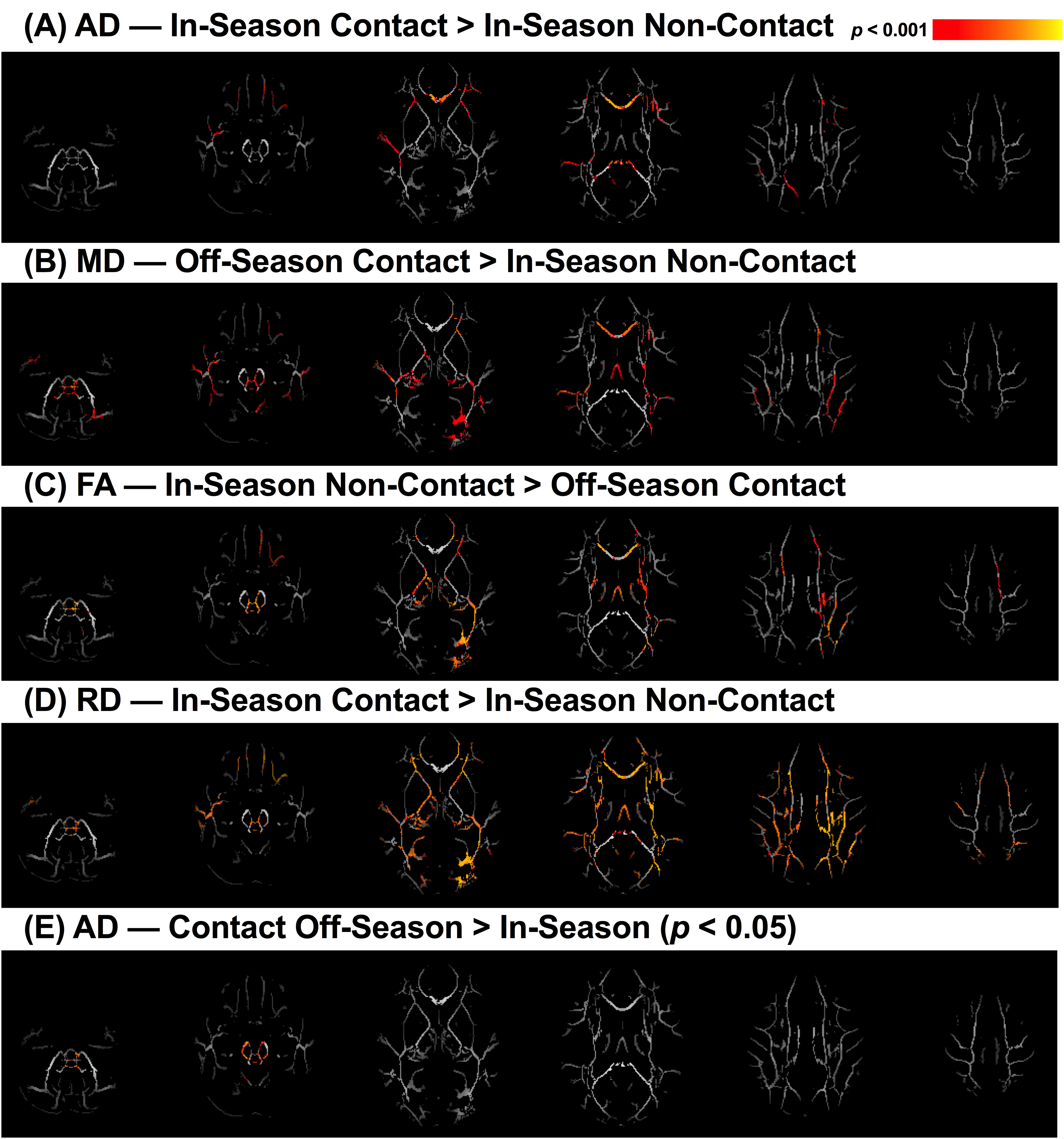
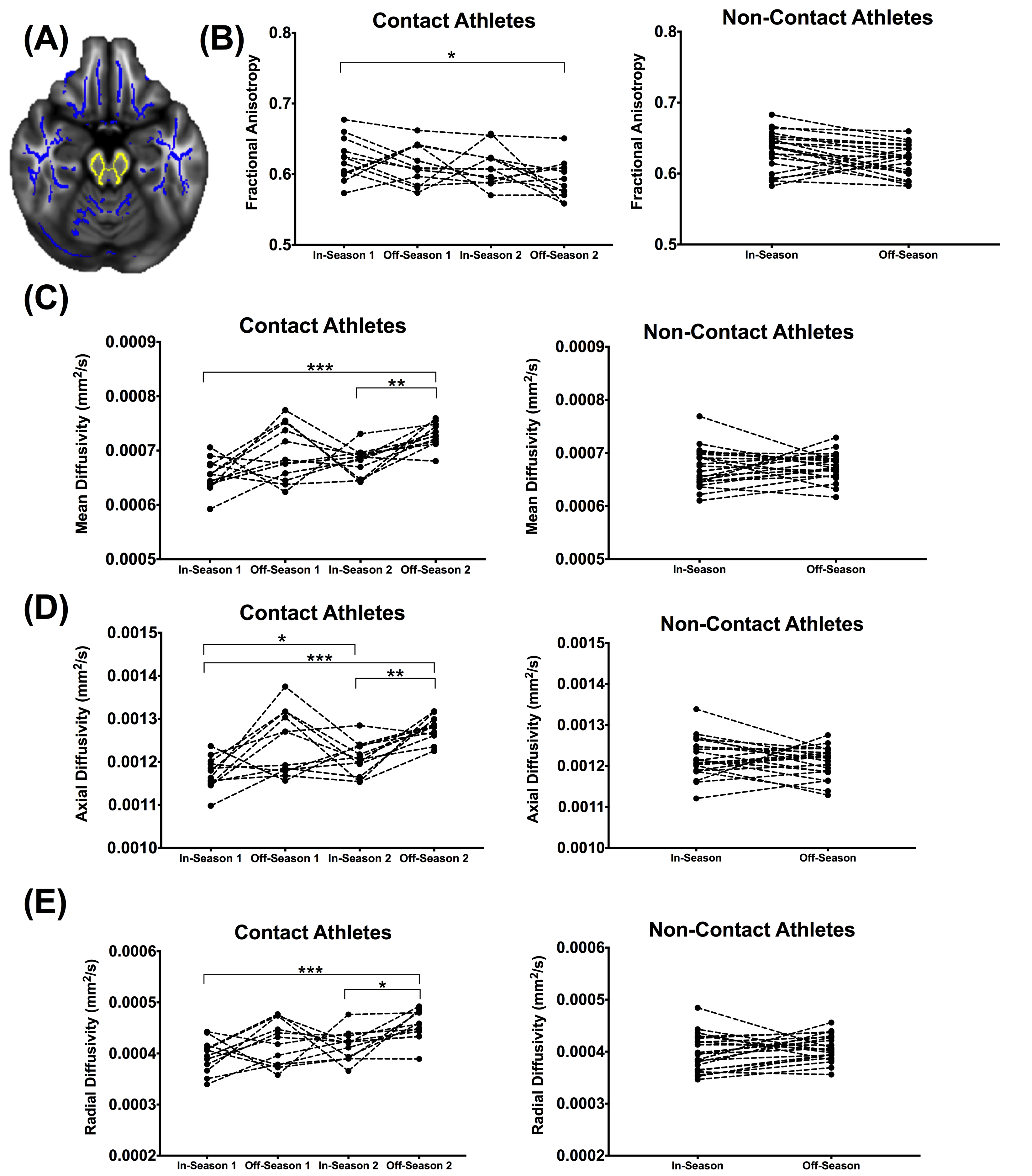
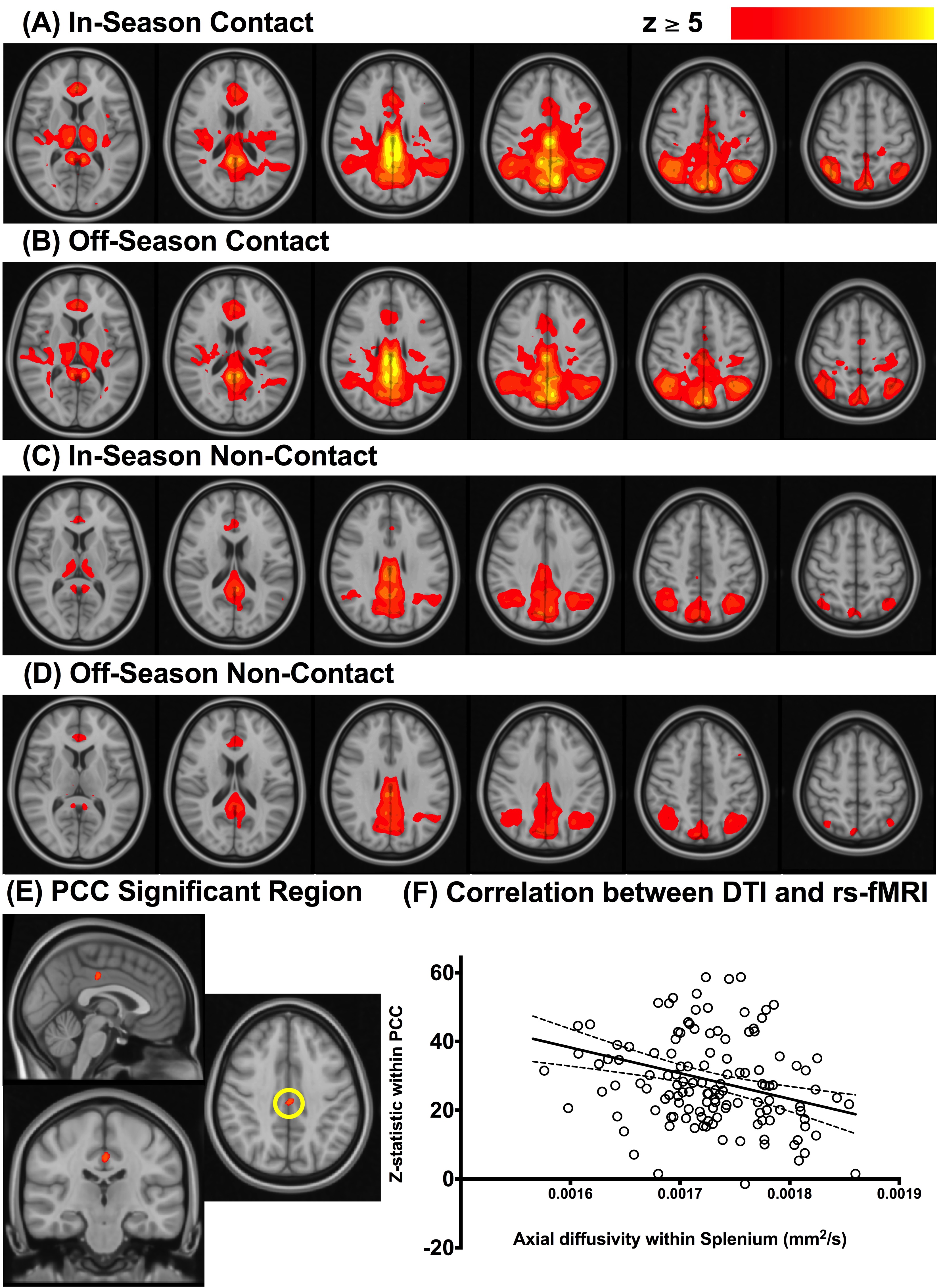
There were significant pairwise improvements in SCAT3 immediate memory and concentration subtest scores in the off-season compared to in-season in contact athletes only (p = 0.01 and 0.003, respectively). Contact athletes on average experienced at least one impact (> 15 g) during practice and game play. Contact athletes had increased MD, RD, and AD and decreased FA (p< 0.001) compared to non-contact athletes (Figure 1). Three segments of the corpus callosum and white matter within the brainstem were identified as regions of interest (ROI). The brainstem ROI had significant pairwise changes in all DTI measures in contact athletes only (Figure 2, p< 0.001) and a significant time*group interaction for AD (t = -3.0, p= 0.003). The AD within the corpus callosum was significantly lower for athletes with a concussion history compared to those without (p< 0.05). Contact athletes had increased connectivity between the posterior cingulate cortex and the DMN and correlated significantly with AD in the splenium of the corpus callosum (Figure 3, r = -0.32, p= 0.0002).Discussion
In this study we observed significantly altered microstructural and functional MRI measures in contact athletes compared to non-contact athletes despite a lack of concussion or symptoms. While fluctuating DTI measures within the brainstem of contact athletes likely reflect an inflammatory response during periods of exposure to subclinical impacts, DTI and rs-fMRI measures in the corpus callosum were significantly altered at both time points compared to non-contact controls suggesting a cumulative effect from years of contact play, with AD being the only measure related to a history of concussion. Our observations may reflect ongoing compensatory mechanisms that help protect the brain from injury and damage due to repetitive subclinical impacts.Conclusion
The biological implication of these findings remains an open question, and in particular to what degree these changes reflect compensatory, reparative or degenerative processes. However based on these results, future studies of concussed athletes must be cognisant when comparing to control groups of contact athletes who experience sports-related impacts and these progressive effects on brain microstructure and function.Acknowledgements
No acknowledgement found.References
1. Breedlove KM, Breedlove EL, Robinson M, et al. Detecting Neurocognitive and Neurophysiological Changes as a Result of Subconcussive Blows in High School Football Athletes. Athl Train Sport Heal Care. 2014; 6(1): 1-9. 2.
2. Shuttleworth-Edwards AB, Smith I, Radloff SE. Neurocognitive vulnerability amongst university rugby players versus noncontact sport controls. J Clin Exp Neuropsychol. 2008; 30(8): 870-884.
3. Killam C, Cautin RL, Santucci AC. Assessing the enduring residual neuropsychological effects of head trauma in college athletes who participate in contact sports. Arch Clin Neuropsychol. 2005; 20(5): 599-611.
4. Abbas K, Shenk TE, Poole VN, et al. Alteration of default mode network in high school football athletes due to repetitive subconcussive mild traumatic brain injury: a resting-state functional magnetic resonance imaging study. Brain Connect. 2015; 5(2): 91-101.
5. McAllister TW, Ford JC, Flashman LA, et al. Effect of head impacts on diffusivity measures in a cohort of collegiate contact sport athletes. Neurology. 2014; 82(1): 63-69.
6. SCAT3. Br J Sport Med. 2013; 47(5):259.
7. Smith, S.M., Jenkinson, M., Johansen-Berg, et al. Tract-based spatial statistics: Voxelwise analysis of multi-subject diffusion data. Neuroimage. 2006; 31: 1487–1505.
8. Griffanti, L., Douaud, G., Bijsterbosch, J., et al. Hand classification of fMRI ICA noise components. Neuroimage. 2017; 154: 188–205.
9. Beckmann, C.F., DeLuca, M., Devlin, J.T., et al. Investigations into resting-state connectivity using independent component analysis. Philos. Trans. R. Soc. B Biol. Sci. 2005; 360: 1001–1013.
Figures