2995
Metabolite Levels Differ in Contact and Non-Contact Sport Female Varsity Athletes1Medical Biophysics, Robarts Research Institute, London, ON, Canada, 2Robarts Research Institute, London, ON, Canada, 3Fowler Kennedy Sport Medicine Clinic, London, ON, Canada, 4University of Western Ontario, London, ON, Canada, 5London Health Sciences Centre, London, ON, Canada
Synopsis
Reduced glutamine levels were previously found in the prefrontal white matter of female varsity rugby athletes after a season of play potentially induced by exercise or caused by sub-concussive hits. The current study examined a group of non-contact female varsity athletes and found no changes in glutamine levels, ruling out an exercise effect. Additionally, differences in absolute N-acetyl aspartate, creatine, myo-inositol, glutamate and glutamine were found between rugby players and non-contact athletes. With the future addition of a sedentary group, these data have the potential to elucidate the beneficial and negative effects of exercise and contact play.
Purpose
Athletes participating in contact sports have a high risk of sustaining a concussion, which can lead to structural and metabolic changes in the brain. Our group previously found reduced glutamine levels in the prefrontal white matter of female varsity rugby athletes at the end of a sports season and after concussion.[1] However, this study was limited by the absence of a non-contact control group to rule out exercise as a cause for the observed metabolite level change. Therefore, the purpose of this study was to quantify changes in brain metabolite levels in female athletes engaged in non-contact sports over the course of a sports season to compare to female athletes who participate in contact sports. It was hypothesized that MRI brain metabolite levels would not significantly change in the non-contact group.Methods
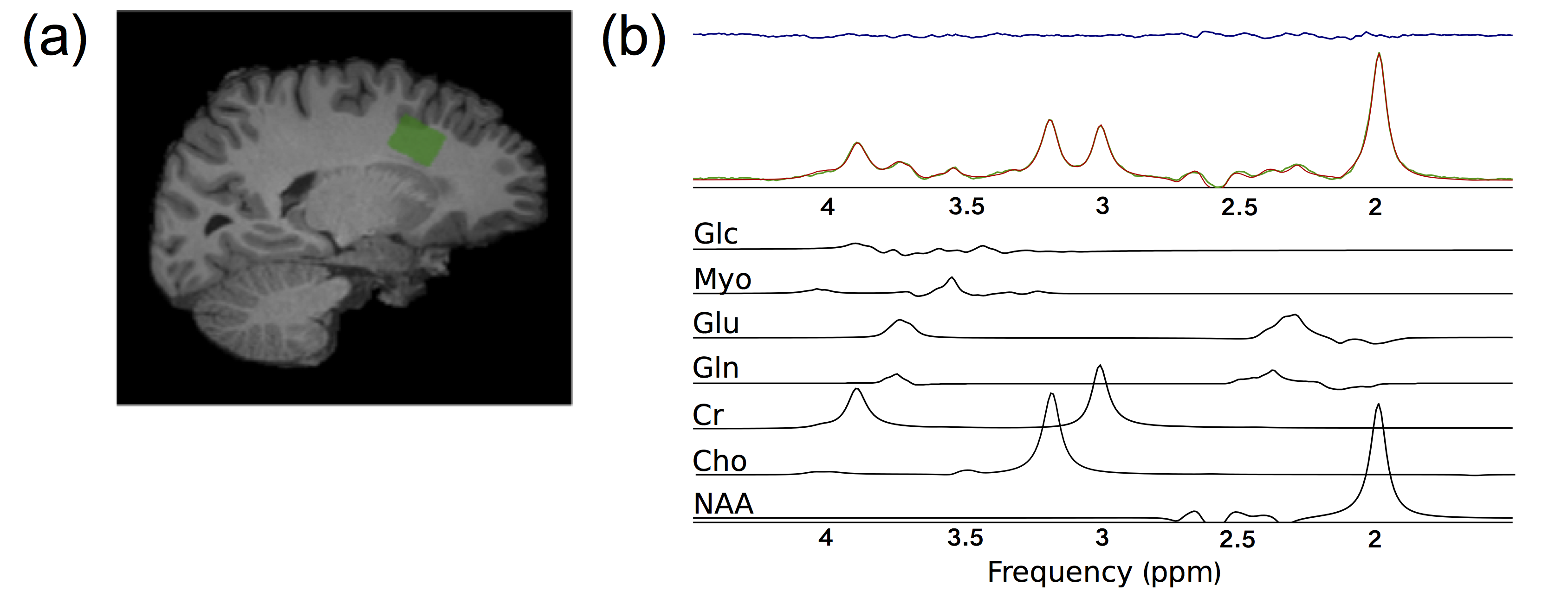
All participants in this study were athletes between the ages of 16 and 22 recruited from women’s varsity rugby, rowing and swim teams. Non-contact (rowers and swimmers, n=31) athletes were scanned at the beginning of the season (in-season) and followed up at the end of the season (off-season) using a Siemens 3T MRI scanner (Erlangen, Germany) using the identical methodology as that previously used for the contact (rugby, n=54) group.[1] 1H magnetic resonance spectroscopy was acquired (TE/TR=135/2000 ms, dwell time = 833 s, number of points = 1024, number of averages suppressed/unsuppressed = 192/8) in the left prefrontal white matter (Fig. 1A). A T2-weighted FLAIR image was acquired to guide the 2 x 2 x 1.5 cm3 voxel placement, and a T1-weighted MPRAGE image was acquired to quantify the GM/WM/CSF composition of the voxel.
The analysis software (fitMAN)[2] created in our laboratory in the IDL (version 5.4 Research Systems Inc., Boulder, CO, USA) programming language was used for analysis. Spectra were lineshape corrected by QUECC[3] then fitted in the time domain using a Levenberg-Marquardt minimization routine (Fig. 1B), using a prior knowledge template that models the metabolite lineshapes acquired from in vitro spectra obtained from aqueous solutions of all metabolites at pH=7.0 prior to the study.[2]
All data analysis was performed using GraphPad Prism version 7.0 for Mac OS X (San Diego, CA). A repeated-measures two-way ANOVA with an alpha value of 0.05 was used to compare metabolites between the in-season and off-season time points between the rowers and swimmers prior to combining both teams into the non-contact group. Then a repeated-measures two-way ANOVA was used to analyze the contact and non-contact groups comparing the in-season and off-season values, alpha value = 0.05.
Results
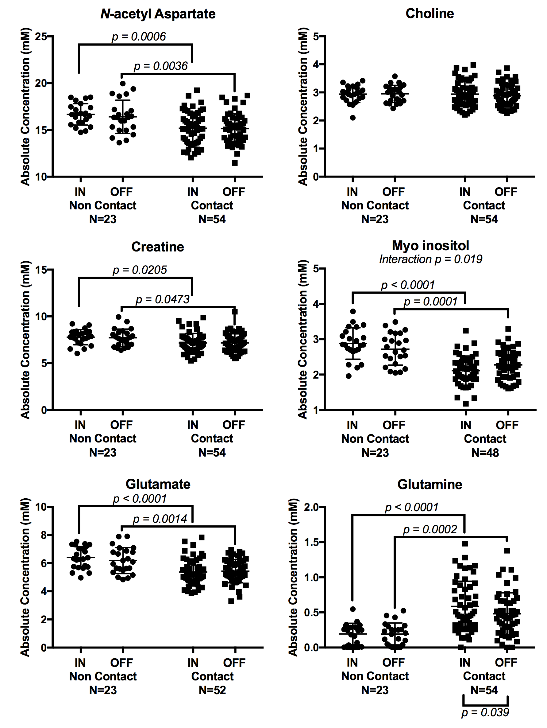
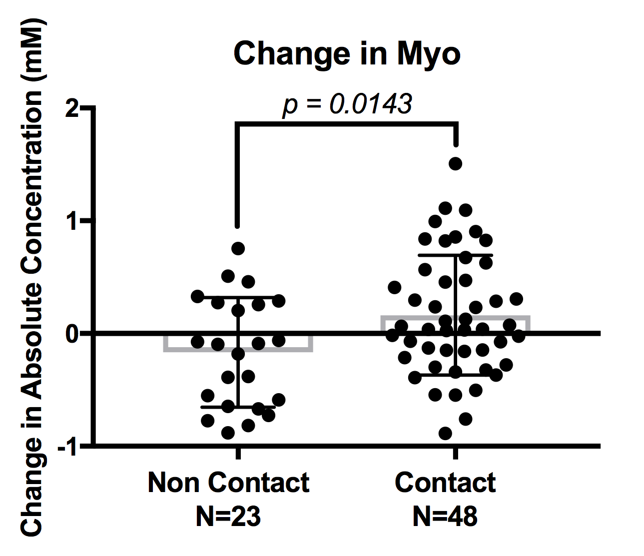
No significant differences were found between the rowers and swimmers so these data were combined into a single non-contact group. Furthermore, there were no metabolite levels including glutamine that significantly changed between the in-season and off-season in the non-contact athletes. However, significant differences between the contact and non-contact groups (p<0.05) were observed for N-acetyl aspartate, creatine, myo-inositol, glutamate and glutamine (Fig. 2). Additionally, a significant interaction (p = 0.019) was observed with myo-inositol. Further analysis revealed a significant difference (p = 0.014) between the change in myo-inositol from in-season to off-season between the contact and non-contact athletes (Fig. 3).Discussion and Conclusion
The current study did not find any change in glutamine levels in non-contact athletes over a season. Therefore, the previously reported reduced glutamine levels in the prefrontal white matter of female varsity rugby athletes at the end of a sports season[1] is unlikely due to an exercise effect since this group of non-contact athletes were matched for activity level. Ruling out exercise, the previous findings in contact-athletes could be interpreted to be the result of concussion history or cumulative sub-concussive impacts over the course of the season.
Reduced N-acetyl aspartate and glutamate levels were observed in the contact group at both in- and off-season time points. These results are in line with previous reports of reduced NAA/Cr in non-concussed female hockey players[4] and reduced glutamate levels in former athletes with a history of concussion.[5] We also observed different directions in myo-inositol change between non-contact and contact groups. Interestingly, increased Myo/Cr has been reported in older athletes with a history of concussion.[6]
Future work includes obtaining data from a non-athlete group to further aid in interpretation of these results. With the comparison of a sedentary group to our non-contact and contact athletes, it may be possible to further elucidate the benefits of activity level on brain biochemistry, as well as the potential negative effects of concussion history and cumulative impacts from contact sports.
Acknowledgements
We would like to thank all the athletes for participating in this study and for the support provided by the coaches, trainers, physicians, and MRI technicians. This work was supported by the Schulich School of Medicine and Dentistry, University of Western Ontario, as well as Brain Canada and the Canada First Research Excellence Fund.References
- Schranz AL, Manning KY, Dekaban GA, Fischer L, Jevremovic T, Blackney K, et al. Reduced brain glutamine in female varsity rugby athletes after concussion and in non-concussed athletes after a season of play. Hum Brain Mapp. 2018;39(4):1489-1499.
- Bartha R, Drost DJ, Williamson PC. Factors affecting the quantification of short echo in-vivo 1H MR spectra: prior knowledge, peak elimination, and filtering. NMR Biomed. 1999;12: 205-216.
- Bartha R, Drost DJ, Menon RS, Williamson PC. Spectroscopic lineshape correction by QUECC: combined QUALITY deconvolution and eddy current correction. Magn Reson Med. 2000;44: 641-645.
- Chamard E, Théoret H, Skopelja EN, Forwell LA, Johnson AM, Echlin PS. A prospective study of physician-observed concussion during a varsity university hockey season: metabolic changes in ice hockey players. Part 4 of 4. Neurosurg. Focus. 2012;33(E4): 1–7.
- De Beaumont L, Tremblay S, Henry LC, Poirier J, Lassonde M, Théoret H, Motor system alterations in retired former athletes: the role of aging and concussion history. BMC Neurol. 2013;13:109.
- Tremblay S, De Beaumont L, Henry LC, Boulanger Y, Evans AC, Bourgouin P, et al. Sports concussions and aging: a neuroimaging investigation. Cereb. Cortex 2012;23:
Figures