2990
Non-invasive Deep-brain Optogenetic fMRI Mediated by 808 nm Infrared-sensitized Upconversion Nanoparticles1Electrical and electronic engineering, University of Hong Kong, Hong Kong, Hong Kong, 2Laboratory of Biomedical Imaging and Signal Processing, The University of Hong Kong, Hong Kong, Hong Kong, 3Department of Mechanical and Biomedical Engineering, City University of Hong Kong, Hong Kong, Hong Kong, 4Department of Biomedical Engineering, Chinese University of Hong Kong, Hong Kong, Hong Kong
Synopsis
Typically, optogenetic fMRI is presented at the target region through an implanted optical fiber. Despite the use of small fibers that range from 100µm-400µm in diameter that will ensure minimal brain tissue injury during insertion, it remains an invasive procedure as small brain regions could be easily damaged. In this study, we aim to demonstrate a solution to make non-invasive optogenetic stimulation viable, particularly when used in combination with fMRI to stimulate deep brain regions. We propose the use of our recently developed upconversion nanoparticles (UCNPs), which can be triggered to emit blue light by penetrative near-infrared light (NIR; 808nm) and excite channelrhodopsins (ChR2) expressed in ventral posteromedial (VPM) thalamocortical excitatory neurons.
Introduction
Optogenetics, a light-sensitive neuromodulation tool that can provide spatiotemporally precise manipulation of genetically defined neuronal populations in vivo, has been widely used in both preclinical basic neuroscience and neuroimaging research. In particular, the combination of optogenetics with blood-oxygen-level-dependent functional MRI (BOLD fMRI) has revolutionized the way complex, large-scale brain networks and their functions are studied1,2.
Typically, optogenetic stimulation is presented at the target region through an implanted optical fiber. Despite the use of small fibers that range from 100µm-400µm in diameter that will ensure minimal brain tissue injury during insertion, it remains an invasive procedure as small brain regions could be easily damaged. Ideally, we sought a setup that can enable optogenetic stimulation without an insertion of fiber into brain tissue. In fact, advances have been made in the form of introducing red-shifted optogenetic actuators/opsins3. Light that is red-shifted has better penetrative capabilities, hence, stimulation can be achieved further away from the target region. However, red-shifted light (550-650nm) is unable to reach deep brain regions below the cortex (~1mm). Moreover, even if the light could sufficiently reach deep brain regions, precision of stimulation remains an issue as light diverges the further it travels.
In this study, we aim to demonstrate a solution to make non-invasive optogenetic stimulation viable, particularly when used in combination with fMRI. Note that the light intensity - an important consideration - required for optogenetic fMRI experiments is higher when compared to electrophysiology studies (40mW/mm2 vs. 5mW/mm2), given the nature of BOLD sensitivity4. Here, we document the use of our recently developed upconversion nanoparticles (UCNPs)5, which can be triggered to emit blue light by penetrative near-infrared light (NIR; 808nm), to excite channelrhodopsins (ChR2) expressed in ventral posteromedial (VPM) thalamocortical excitatory neurons.
Methods
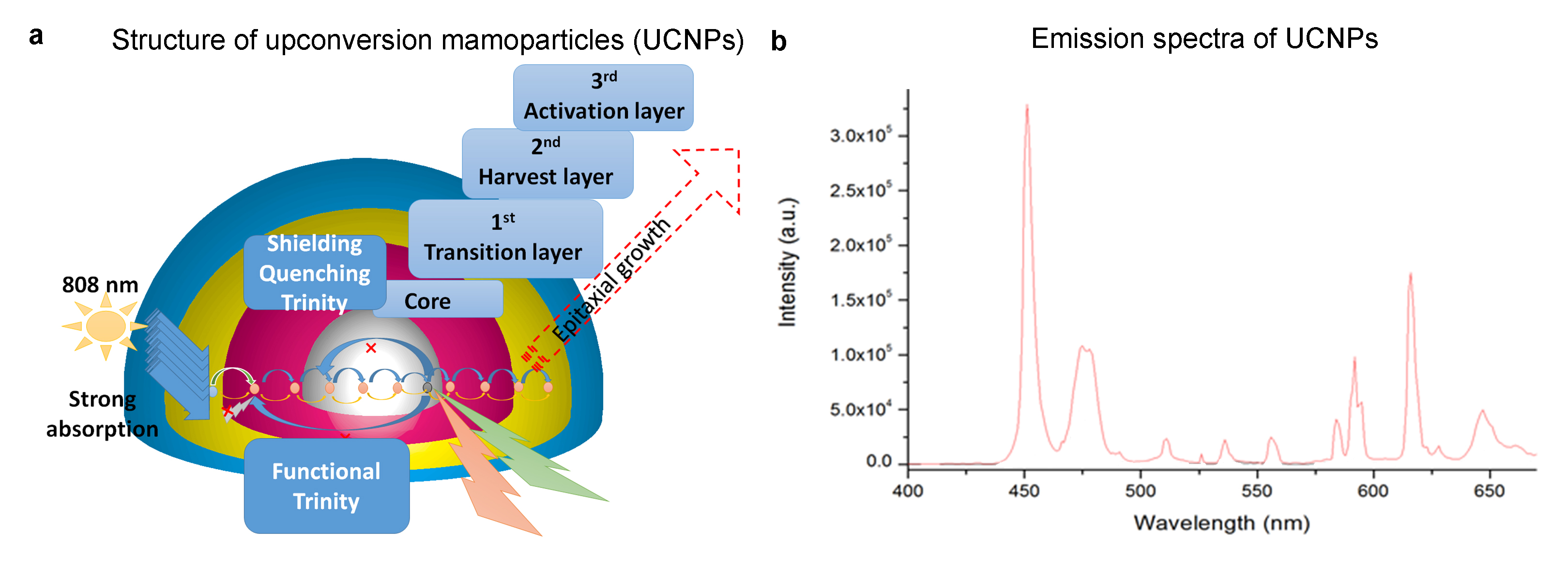
Silica-coated UCNPs synthesis and characterization: The core-multishells UCNPs are composed of the NaGdF4:Yb49%,Tm1% core, which was first synthesized through thermal decomposition; by coating with a three layers name as “Nd3+-Trinity system” to enhance its luminescence property5. Subsequently, UCNPs were coated with silica to minimize cytotoxicity through sol-gel process (Figure 1a). Subsequently, the yield product was collected by centrifugation at 6,000rpm. The emission spectra showed that blue light ~450nm is predominantly emitted by UCNPs when exposed to NIR (Figure 1b).
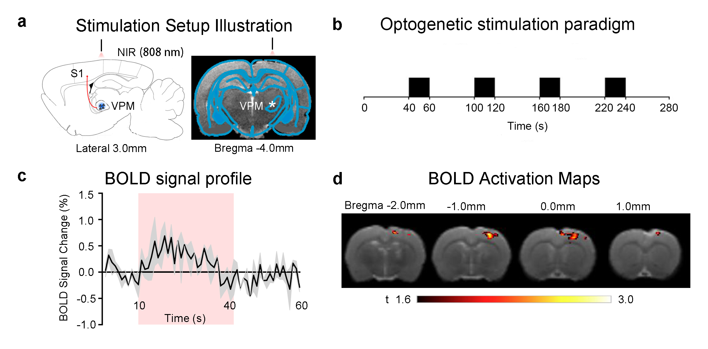
Animal preparation and UCNPs mediated optogenetic stimulation: 3μl AAV5-CaMKIIα::ChR2(H134R)-mCherry was injected at VPM of SD rats (n=4, 200-250g, male). Four weeks after injection, 0.5µl UCNPs was deposited at VPM. All experiments were performed under 1.0% isoflurane. 808nm NIR light was presented at 10Hz with a 20s on 40s off block design paradigm, and 4 pulses, 0.5s duration at 8Hz once every 30s (10% duty cycle, 400mW/mm2, 2mm away from cortex; Figure 2a).
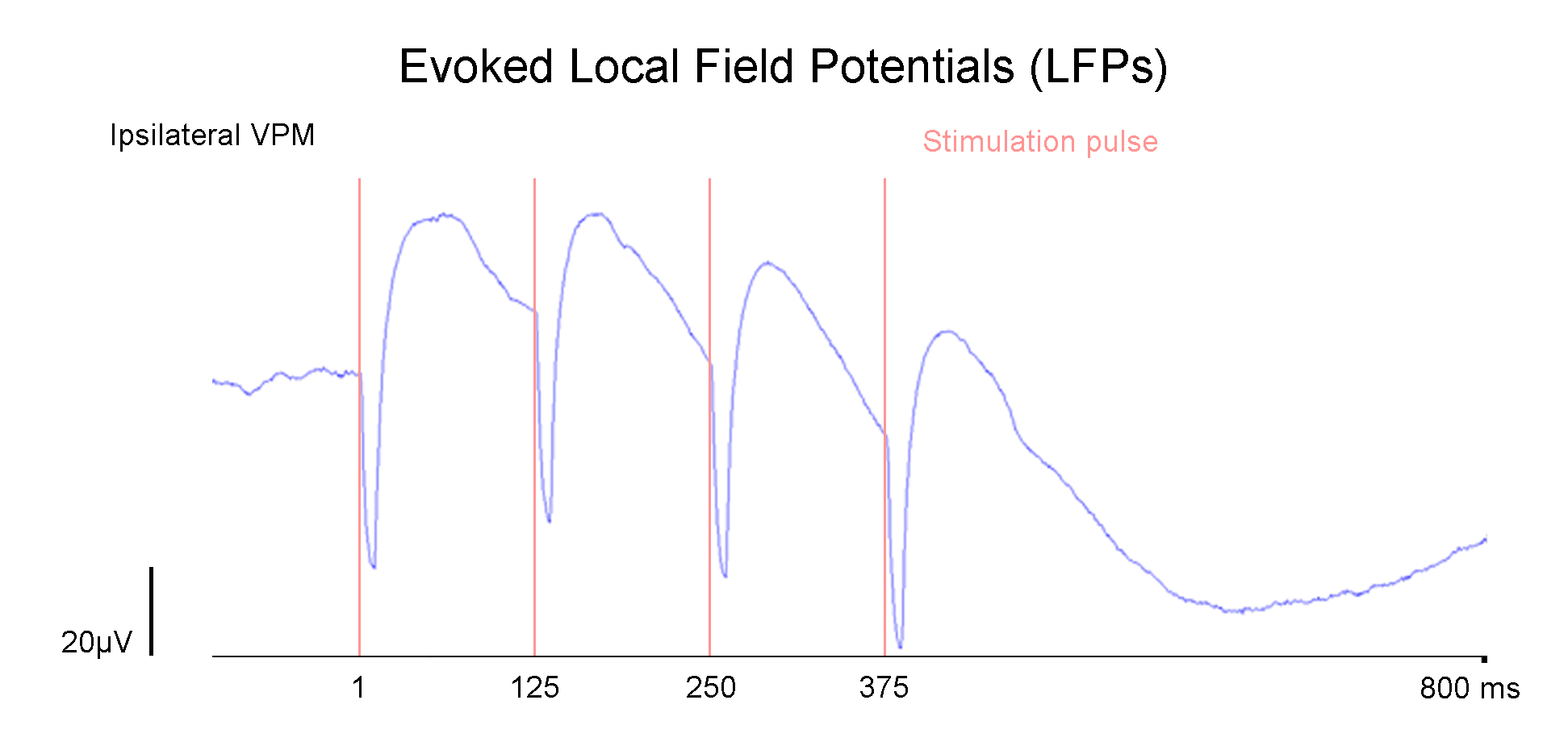
fMRI and electrophysiology experiments and data analyses: fMRI data were acquired at 7T using GE-EPI (FOV=32×32mm2, matrix=64×64, α=56°, TE/TR=20/1000ms, and 16 contiguous slices with 1mm thickness). Data were preprocessed before coherence analysis was applied to identify significant BOLD responses (P<0.001). Evoked local field potentials (LFPs; n=2) were acquired at ipsilateral somatosensory cortex (S1) using multi-depth electrodes (16 channels). LFP data were digitized at 1kHz and bandpass-filtered (0.1Hz-150Hz).
Results
UCNPs mediate excitation of ChR2 at VPM to activate the thalamo-cortical circuit: Using VPM as a model, given its well-defined thalamo-cortical projections to S16, we observed activations in ipsilateral S1 upon exposure of UCNPs to the NIR light (Figure 2b, c). This finding demonstrates that UCNPs can be triggered at deep brain regions, have high fidelity when exposed to NIR and emit sufficient light intensity to excite ChR2; leading to detectable BOLD activations. Subsequent electrophysiology recordings conducted in ipsilateral VPM at similar conditions as the fMRI experiments revealed evoked LFPs that followed the presentation of NIR light. (Figure 3).
Discussion and Conclusion
Our work demonstrates the in vivo feasibility of the implementation of the UCNPs as an actuator in optogenetic functional MRI experiments. However, future developments would still need to be made to improve the efficiency of light emission from UCNPs when exposed to NIR. In the present study, the activations in ipsilateral S1 is much weaker than that shown in our previous optogenetic fMRI study of the VPM that utilized a fiber approach to deliver light4. Nevertheless, our present study has demonstrated a viable non-invasive strategy that holds great promise in the development of future fiberless optogenetic experiments. Specifically, due to the multiple luminescence capability of UCNPs, they can be deployed for multiplexed stimulation purposes to excite/inhibit individually small, yet closely spaced brain regions that would otherwise be challenging for fiber-based approaches.Acknowledgements
This work was supported by the Hong Kong Research Grant Council (C7048-16G and HKU17103015 to E.X.W.).References
1. Lee, J. H. et al. Global and local fMRI signals driven by neurons defined optogenetically by type and wiring. Nature 465, 788-792, doi:10.1038/nature09108 (2010).
2. Chan, R. W. et al. Low-frequency hippocampal-cortical activity drives brain-wide resting-state functional MRI connectivity. Proc Natl Acad Sci U S A 114, E6972-E6981, doi:10.1073/pnas.1703309114 (2017).
3. Chuong, A. S. et al. Noninvasive optical inhibition with a red-shifted microbial rhodopsin. Nat Neurosci 17, 1123-1129, doi:10.1038/nn.3752 (2014).
4. Leong, A. T. et al. Long-range projections coordinate distributed brain-wide neural activity with a specific spatiotemporal profile. Proc Natl Acad Sci U S A 113, E8306-E8315, doi:10.1073/pnas.1616361113 (2016).
5. Guo, S., Tsang, M.-K., Lo, W.-S., Hao, J. & Wong, W.-T. 808 nm excited energy migration upconversion nanoparticles driven by a Nd3+–Trinity system with color-tunability and superior luminescence properties. Nanoscale 10, 2790-2803, doi:10.1039/C7NR07026H (2018).
6. Cruikshank, S. J., Urabe, H., Nurmikko, A. V. & Connors, B. W. Pathway-specific feedforward circuits between thalamus and neocortex revealed by selective optical stimulation of axons. Neuron 65, 230-245, doi:10.1016/j.neuron.2009.12.025 (2010).
Figures